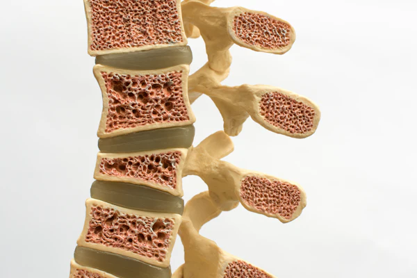
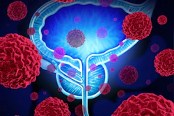
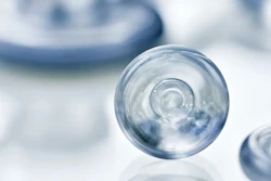
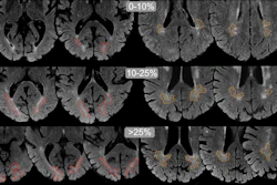
Dr. Perry PickhardtCTAtlas of Gastrointestinal Imaging, Chapter 7 -- The PancreasIntraductal papillary mucinous neoplasm (IPMN) is a distinct subset of MCN that is being recognized with increasing frequency.August 27, 2007CTAtlas of Gastrointestinal Imaging, Chapter 5 -- The Colon and RectumImaging evaluation of the large intestine has undergone remarkable advances over the past decade, particularly with the advent of CT colonography (CTC, also referred to as virtual colonoscopy). Optical colonoscopy (also referred to as conventional or invasive colonoscopy) and CTC allow for complementary diagnostic evaluation of the colonic mucosa.August 27, 2007CTAtlas of Gastrointestinal Imaging, Chapter 4 -- The Mesenteric Small BowelMesenchymal tumors are a heterogeneous group of intramural lesions that arise deep to the mucosa.August 27, 2007HomeAtlas of Gastrointestinal Imaging Figure 1.3.4 IPMN with invasive carcinomaAugust 26, 2007HomeAtlas of Gastrointestinal Imaging Figure 1.3.3 Side branch IPMNAugust 26, 2007HomeAtlas of Gastrointestinal Imaging Figure 1.3.2 Pancreas divisum with dorsal duct IPMNAugust 26, 2007HomeAtlas of Gastrointestinal Imaging Figure 1.3.1 Main duct IPMNAugust 26, 2007HomeAtlas of Gastrointestinal Imaging FIGURE 2.1.7 Rectal fistulas complicating ulcerative colitisAugust 26, 2007HomeAtlas of Gastrointestinal Imaging FIGURE 2.1.6 Dysplasia and carcinoma in long-standing ulcerative colitisAugust 26, 2007HomeAtlas of Gastrointestinal Imaging Figure 2.1.5 Long-Standing ulcerative colitisAugust 26, 2007Page 1 of 3Next PageTop StoriesCTOpportunistic screening can reveal osteoporotic vertebral fracturesA new study demonstrates the potential of opportunistic screening in improving osteoporosis care.MRIBlood-based testing could improve prostate cancer screening with MRIImage ProcessingAdvancing precision medicine through quantitative imagingMRI"Dirty" brain white matter shows no link to dementia in older adultsMolecular ImagingNew PET tracer shows promise for guiding pancreatic cancer therapy