


More in Home
Could more imaging access improve prison cancer care?
October 15, 2025


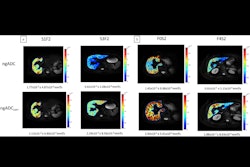
qHDMI differentiates radial scars from invasive breast cancer
October 15, 2025

Lung biopsy before surgery linked to cancer recurrence
October 14, 2025


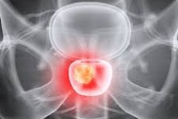
PSMA-PET/CT reporting varies widely in the U.S.
October 14, 2025


