PET/CT imaging has revealed brain regions in leukemia patients with lower glucose metabolism after chemotherapy, according to a study published October 16 in the Journal of Nuclear Medicine.
The findings support research into metabolic imaging biomarkers for early detection and intervention in patients experiencing cognitive impairment, noted first author Ahmed Msherghi, MD, of the MD Anderson Cancer Center in Houston, TX, and colleagues.
“The use of F-18 FDG-PET/CT imaging to monitor brain metabolism in chemotherapy patients may help identify vulnerable populations and guide personalized treatment plans,” the group wrote.
Chemotherapy-induced cognitive impairment (CICI), also referred to as chemo brain, is a common neurologic condition due to anticancer medications, the authors explained. It is a subtype of cancer-related cognitive impairment that specifically results from systemic chemotherapy. The condition affects memory, attention, and executive function, and typically lasts for six to nine months after treatment.
While previous studies using F-18 FDG-PET/CT have identified distinct patterns of glucose uptake in various brain cortex regions, to date, no studies have investigated the relationship between the timing of chemotherapy and brain glucose metabolism or have explored how the intrathecal route may influence the metabolic effects on the brain, the authors noted.
To fill the knowledge gap, the group retrospectively analyzed data from patients with newly diagnosed or relapsed leukemia who underwent treatment and had undergone PET/CT brain scans (Biograph Vision Quadra, Siemens Healthineers) at their hospital between January 2023 and August 2024.
The researchers included 29 control patients who had not received any chemotherapy, 22 patients who had received chemotherapy more than one year before the scan, and 49 patients who were either currently undergoing chemotherapy or had completed their chemotherapy course within one year before the scan. The researchers quantified regional F-18 FDG radiotracer uptake with MIM software (GE HealthCare) using normalized voxel values and compared findings across the group.
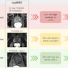
 A visual abstract of the study.Journal of Nuclear Medicine
In patients 55 or older, reduced metabolism was observed in the Rolandic operculum (1.12 vs. 1.19; p < 0.001) and inferior frontal gyrus (1.16 vs. 1.19; p = 0.05). In addition, recent chemotherapy recipients showed increased metabolism in the fusiform gyrus (1.34 vs. 1.27; p = 0.04) and insula, whereas long-term survivors did not. Finally, intrathecal chemotherapy was linked to reduced thalamic metabolism (1.11 vs. 1.15; p = 0.02).
“Chemotherapy is associated with voxel-based brain metabolic alterations, particularly in areas governing cognition and emotion. Effects are more pronounced in older adults and those receiving intrathecal treatment,” the group wrote.
Ultimately, future studies should be designed as prospective trials with sufficient statistical power to validate these correlations and should include patients across different cancer types and treatment regimens, as well as assessments over time to better understand the condition, the researchers suggested.
“These findings support research into metabolic imaging biomarkers for early detection and intervention in chemotherapy-induced cognitive impairment,” they concluded.
The full study is available here.