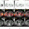
| FIGURE 1.1.4 Flat adenomas 3D endoluminal CTC image (A) and 2D CTC image with soft tissue windowing (B) show a slightly raised, flat 7-mm tubular adenoma (arrowheads) adjacent to the rectal catheter. 3D endoluminal CTC image (C) and corresponding colonoscopy image (D) from a second patient show a similar-appearing 6-mm, flat tubular adenoma (C, arrowheads) in the sigmoid colon. 3D endoluminal CTC (E) and corresponding colonoscopy (F) images from a third patient show a 3.5-cm tubulovillous adenoma with a broad elongated appearance. This lesion could be classified as flat or sessile, depending on the specific definition used. (A and B from Pickhardt PJ, Nugent PA, Mysliwiec PA, et al: Location of adenomas missed at optical colonoscopy. Ann Intern Med 2004; 141:352-359; C and D from Pickhardt PJ, Nugent PA, Choi JR, Schindler WR: Flat colorectal lesions in asymptomatic adults: Implications for screening with CT virtual colonoscopy. AJR 2004; 183:1343-1347.) |
Atlas of Gastrointestinal Imaging Figure 1.1.4 Flat adenomas
Latest in Home