Forensic CT helps medical examiners determine cause of death -- and thus avoid autopsy, a procedure that can not only be upsetting for a decedent's loved ones but also time-consuming for medical examiner staff, according to Kalpana Kanal, PhD, of the University of Washington (UW) Medical Center in Seattle.
"[Someone asked me], 'Do you see radiologists showing up in a CSI show?'" she told AuntMinnie in a video interview. "Heck yeah, we could have a radiology CSI, with the medical examiner doing their part and radiology helping to determine whether an autopsy is needed during daily rounds."
Kanal is a medical physicist, UW's section chief of diagnostic physics, and a contributing developer of a forensic CT program in Seattle. Along with partners J. Matthew Lacy, MD, chief medical examiner at King County's medical examiner's office; Harborview Medical Center's director of radiology, Jonathan Medverd, MD; and fellow UW medical physicist David Zamora, Kanal helped launch the program three years ago. Initially, it used a CT unit at UW, but in May of this year, the examiner's office installed its own CT scanner. UW radiologists read the exams.
"[The program was launched] in response to the question: How can we leverage imaging to help with death investigations?" she said. "We see this as a service to our community, helping families who don't want their loved ones to go through an autopsy to understand cause of death in a noninvasive way."
In the interview, Kanal discussed how exactly CT is used for this indication, the process of obtaining and interpreting the exams, and what obstacles to further implementation forensic CT faces.
Using CT to determine cause of death is a helpful way to reduce the workload of the medical examiner's office as well, Kanal said, citing a recent study she and colleagues published in the Journal of Computer Assisted Tomography, which found that in 45% of cases, there was no need for autopsy because CT provided the answers needed.
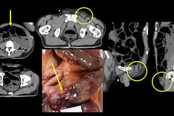
 This image depicts comminuted skull fractures sustained in a pedestrian motor vehicle accident. These are difficult to document by a standard autopsy because the individual fragments are displaced during the internal examination and dissection of the head.Kalpana Kanal, PhD.
In the interview, Kanal outlined a typical forensic CT protocol:
She acknowledged that one of forensic CT's big hurdles is that it is not currently reimbursed: Radiologists who read these exams are essentially volunteers.
Nevertheless, radiologists can play a key role in this particular use of CT imaging that can streamline and improve the process of determining an individual's cause of death, she concluded.