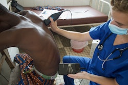
Transport team members can use point-of-care ultrasound (POCUS) to obtain cardiac images in infants with congenital heart disease (CHD), suggest findings published October 18 in Air Medical Journal.
A group led by Brittany Shutes, MD, from Nationwide Children’s Hospital in Columbus, OH, found that after training, transport team members could acquire sufficient quality POCUS images that allow for identifying and triaging CHD in infants who present with hypoxemia.
“This novel, resource-conscious application has the potential to streamline the care of hypoxemic newborns after undergoing protocol efficiency refinement, independent proficiency testing, and study of remote application,” Shutes and colleagues wrote.
Given the current limitations of prenatal CHD assessment, neonatal transport team members may be on the front line for assessing critical CHD in infants via ultrasound. They also may have more responsibility for guiding therapies and transporting infants to appropriate care locations.
Prior research suggests that effective training can build competence and confidence among non-sonographers who use POCUS. However, the researchers noted a lack of data on the potential use of a hybrid patient care model that combines targeted, locally acquired ultrasound images with real-time, remote interpretation by pediatric cardiologists.
Shutes and colleagues evaluated the feasibility of training transport team members in focused cardiac ultrasound of newborns. They also studied the quality of cardiac images provided by a handheld ultrasound device and the accuracy of the diagnosis of CHD in infants.
An overview of the focused cardiac ultrasound protocol described in the study. Images are republished under a Creative Commons license (CC BY 4.0).Elsevier
In the first phase, the investigators surveyed transport team members on their prior experience with ultrasound. From there, the transport team members underwent structured education and hands-on simulation sessions on cardiac anatomy and ultrasound. They completed knowledge assessments afterward.
For the second phase, the participants then performed supervised bedside ultrasound on hospitalized infants to practice skills, familiarize themselves with the POCUS device, and learn the focused cardiac ultrasound protocol. Shutes and colleagues then compared the ultrasound images obtained by the participants with comprehensive images assessed by two pediatric cardiologists.
Final analysis included data collected from 25 participants from phase I and 17 participants from phase II. The participants significantly improved their skills, with median scores rising from 47% before training to 79% post-training (p < 0.05).
In phase II, 17 transport members completed 42 scans on 14 unique infants. Of these, 10 had confirmed CHD. Of the total scans, 39 had sufficient image quality to make a diagnosis, with 37 having “high” diagnostic certainty and 36 matching complete transthoracic echocardiograms with no missed diagnosis.
Finally, the study authors reported a median completion time of 17 minutes, with a range of 12.8 to 20.6 minutes.
They suggested that further refinement and application of this approach could shorten the time it takes for a hypoxemic newborn with undiagnosed critical CHD to receive appropriate care.
“The ultimate goal in future implementation would be to provide another piece of clinical information in a timely fashion to help in triaging patient destination and targeted therapies,” the group concluded. “The next phase of this research should include testing of independent proficiency of transport team personnel.”
Read the entire study here.



![Examples of ultrasound findings and techniques. (A) Images in a 39-year-old male patient with a mass in the left thigh. The mass is heterogeneous on the B-mode US image (compared with the patient in D) and showed increased microvascularity (superb microvascular imaging [SMI]) and shear-wave elastography (SWE) values. Undifferentiated pleomorphic sarcoma was diagnosed at biopsy (with pleomorphic rhabdomyosarcoma in surgical specimen). (B) Images in an 18-year-old male patient with a mass in the left leg. The mass is hypoechoic on the B-mode image, with no other findings suggestive of malignancy. The lesion is in contact with the cortex of the tibia, which is slightly irregular. CT revealed a doubtful anteromedial tibial erosion. The microvascular study demonstrated high vascularization, suggestive of malignancy. Periosteal Ewing sarcoma was diagnosed with both histologic and immunohistochemical confirmation. (C) Images in a 69-year-old female patient with a lump growing on the outside of the left leg. Multiple SWE examinations were performed (please note the high values obtained in the measurements, whereas the color map highlights the stiffness relative to adjacent tissues). SMI showed areas of increased vascularization to target for sampling. Undifferentiated spindle cell sarcoma was diagnosed at biopsy, with residual leiomyosarcoma in the surgical specimen after neoadjuvant therapy. (D) Images in a 56-year-old female patient with a mass in the right thigh. The mass is heterogeneous at both B-mode ultrasound (similar to patient A) and MRI (coronal T2-weighted spectral attenuated inversion recovery [SPAIR]; T1-weighted pre-contrast and postcontrast imaging), which even shows uptake after the administration of paramagnetic contrast material, which is traditionally suggestive of malignancy. Low values at SMI and elastography are suggestive of benignity. Spindle cell lipoma was diagnosed at biopsy, with atypical spindle cell lipomatous tumor in the surgical specimen.](https://img.auntminnie.com/mindful/smg/workspaces/default/uploads/2026/08/images-radiol250278fig2.APCFLSvX6p.jpg?auto=format%2Ccompress&fit=crop&h=100&q=70&w=100)


![Examples of ultrasound findings and techniques. (A) Images in a 39-year-old male patient with a mass in the left thigh. The mass is heterogeneous on the B-mode US image (compared with the patient in D) and showed increased microvascularity (superb microvascular imaging [SMI]) and shear-wave elastography (SWE) values. Undifferentiated pleomorphic sarcoma was diagnosed at biopsy (with pleomorphic rhabdomyosarcoma in surgical specimen). (B) Images in an 18-year-old male patient with a mass in the left leg. The mass is hypoechoic on the B-mode image, with no other findings suggestive of malignancy. The lesion is in contact with the cortex of the tibia, which is slightly irregular. CT revealed a doubtful anteromedial tibial erosion. The microvascular study demonstrated high vascularization, suggestive of malignancy. Periosteal Ewing sarcoma was diagnosed with both histologic and immunohistochemical confirmation. (C) Images in a 69-year-old female patient with a lump growing on the outside of the left leg. Multiple SWE examinations were performed (please note the high values obtained in the measurements, whereas the color map highlights the stiffness relative to adjacent tissues). SMI showed areas of increased vascularization to target for sampling. Undifferentiated spindle cell sarcoma was diagnosed at biopsy, with residual leiomyosarcoma in the surgical specimen after neoadjuvant therapy. (D) Images in a 56-year-old female patient with a mass in the right thigh. The mass is heterogeneous at both B-mode ultrasound (similar to patient A) and MRI (coronal T2-weighted spectral attenuated inversion recovery [SPAIR]; T1-weighted pre-contrast and postcontrast imaging), which even shows uptake after the administration of paramagnetic contrast material, which is traditionally suggestive of malignancy. Low values at SMI and elastography are suggestive of benignity. Spindle cell lipoma was diagnosed at biopsy, with atypical spindle cell lipomatous tumor in the surgical specimen.](https://img.auntminnie.com/mindful/smg/workspaces/default/uploads/2026/08/images-radiol250278fig2.APCFLSvX6p.jpg?auto=format%2Ccompress&fit=crop&h=112&q=70&w=112)