Growth in its Image-Guided Therapy (IGT) and Ultrasound units helped Philips Healthcare's Diagnosis and Treatment businesses achieve a 5% increase in sales for its fourth quarter. Sales were flat, however, in its Connected Care and Health Informatics businesses.
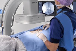
For the period (end-December 31, 2018), Philips reported 2.2 billion euros ($2.51 billion U.S.) in sales for its Diagnosis and Treatment businesses, up 5% on a comparable basis from the 2.09 billion euros ($2.39 billion) in the fourth quarter of 2017. Growth was driven by double-digit gains in IGT and high-single-digit growth in ultrasound. Parent Royal Philips also noted that its Diagnostic Imaging business achieved double-digit growth in order intake.
Overall, the Diagnosis and Treatment businesses had adjusted earnings before interest, taxes, and amortization (EBITA) of 294 million euros ($335.8 million) in the fourth quarter, up from 266 million euros ($303.9 million) in the same period a year ago.
Meanwhile, the vendor's Connected Care and Health Informatics businesses produced 936 million euros ($1.07 billion) in sales, flat on a comparable basis with the 912 million euros ($1.04 billion) reported in the fourth quarter of last year. Low-single-digit growth in its Therapeutic Care and Monitoring and Analytics businesses were offset by a low-single-digit decline in healthcare informatics, according to the vendor.
However, Philips said that it produced double-digit growth in order intake, driven by its Monitoring and Analytics and Health Informatics businesses. Overall, the Connected Care and Health Informatics businesses had adjusted fourth-quarter EBITA of 114 million euros ($130.2 million), down from 169 million euros ($193.1 million) in the fourth quarter of 2017.
Thanks to double-digit growth in IGT and ultrasound, as well as low-single-digit growth in diagnostic imaging, the company's Diagnostic and Treatment businesses had 2018 sales of 7.25 billion euros ($8.28 billion), up 7% on a comparable basis from the 6.89 billion euros ($7.87 billion) reported in 2017. Adjusted EBITA reached 696 million euros ($795.1 million), compared with 543 million euros ($620.3 million) last year.
The firm's Connected Care and Health Informatics businesses generated 3.08 billion euros ($3.52 billion) in sales, flat on a comparable basis from the 3.16 billion euros ($3.61 billion) produced a year ago. The flat sales growth reflected low-single-digit growth in healthcare informatics and flat results in its Monitoring and Analytics and Therapeutic Care businesses, Philips said. The Connected Care and Health Informatics businesses had adjusted EBITA of 341 million euros ($389.6 million), down from 372 million euros ($425 million) reported in 2017.