PHILADELPHIA -- A conversational AI workflow could help improve extended focused assessment with sonography in trauma (eFAST) exams, suggest findings presented May 29 at the American Institute of Ultrasound in Medicine (AIUM) annual meeting.
Emergency sonographers showed improved performance with assistance from ChatGPT-5, comparable to conventional instructor-based training, according to a pilot study presented by Nardine Francis from the California University of Science and Medicine in Colton.
“We found that AI is a feasible operational tool that can be used to help teach eFAST exams in medical education,” Francis told AuntMinnie.
eFAST exams use ultrasound to detect conditions in trauma patients, including intraperitoneal free fluid, pericardial effusion, hemothorax, and pneumothorax. This can be challenging for novice learners since these exams are operator-dependent and time-sensitive.
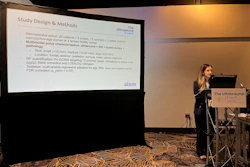
 Nardine Francis from the California University of Science and Medicine presents research at AIUM 2026 showing how ChatGPT could help novice learners in eFAST training.AuntMinnie
Previous research suggests that over three in five U.S. MD schools have ultrasound integration, but only one in five has ultrasound education as a current institutional priority. Faculty bandwidth has been cited as a barrier to ultrasound training, highlighting the need for reproducible coaching.
The Francis team studied whether ChatGPT-5 could effectively assist eFAST practice in novice learners and compared results to those from instructor-based learning.
The pilot study included 16 first-year students at the university who had no prior formal ultrasound training. The students were randomized to either AI-assisted or instructor-assisted practice in a 1:1 ratio. In the AI-assisted group, learners entered typed questions and uploaded still ultrasound images into ChatGPT-5. The chatbot provided feedback limited to probe positioning, scan-window landmarks, image quality, and troubleshooting.
Nardine Francis shares how using conversational AI could address education gaps in eFAST training.
Both AI- and instructor-assisted groups showed improved knowledge and confidence, with both areas being close in scoring.
Comparison between AI-assisted, instructor-assisted eFAST training | |||
Measure | Instructor assistance | AI assistance | P-value |
Baseline knowledge | 30.0% | 32.5% | 0.8 |
Post-training knowledge | 85.0% | 87.5% | 0.7 |
Baseline confidence | 1.54 | 1.40 | 0.4 |
Post-training confidence | 5.25 | 4.98 | 0.2 |
Francis reported the AI workflow being workable, with learners able to interact using typed queries and still images without embedded ultrasound AI algorithms.
Despite the success seen in the study, Francis cautioned that long-term knowledge retention cannot yet be inferred and that the results cannot be generalized to unsupervised clinical use.
She said that future work will focus on AI’s best curricular role, with studies having larger sample sizes among learning cohorts.
“We probably want to see whether AI on its own is good or maybe [serve as] an adjunct with instruction by faculty if it’s even better,” she told AuntMinnie.
For more of AuntMinnie's coverage from AIUM 2026, click here.