
PHILADELPHIA – Ultrasound is a useful imaging tool for global health initiatives, but imagers who lead these health missions must go in with the goal of building capacity for hospitals around the world.
That was the main takeaway from a May 30 presentation at the American Institute of Ultrasound in Medicine (AIUM), where three speakers presented on their experiences working with ultrasound around the world.
“Building capacity is about strengthening systems,” said Yamilé Blain, MD, from the University of Miami in Florida. “It’s about making sure that the infrastructures are solid, that resources are available, and that the end users… are skilled.”
In her talk, Blain shared her experience working with three teaching hospitals in Haiti, where she worked with the Humanitarian Radiology Development (HRD) Corps to deliver ultrasound scanners and training to clinicians. Blain is from Haiti and completed her radiology residency there.
Over a three-year period (2020 to 2022), the hospitals reported these scanners being used in 1,458 female pelvic exams and 1,075 abdominal exams.
Blain said for these global health missions to be successful, leaders need to build capacity for long-term sustainability. These include assessing the needs of hospitals, having extra scanners available in case of equipment malfunctions, effective training, periodic maintenance, and the ability to repair scanners in quick fashion.
“Know your audience. Know what’s needed based on the reality there and not our own reality in practice here [in the U.S.],” Blain said. “For every radiology program’s ultrasound-building capacity, improvement is needed. Pick equipment that makes sense for the place.”
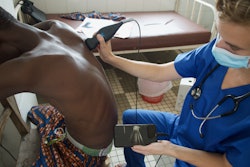
In another presentation, Hunter Wynkoop, MD, from Nationwie Children’s Hospital in Columbus, OH talked about her time working in Sub-Saharan Africa to deliver pediatric ultrasound equipment and training. Here, pediatric mortality and long-term morbidity are affected by sickle cell disease tied to related vasculopathy and stroke, and acute central nervous system infections.
 Hunter Wynkoop, MD, (far left) shares insights from her time working in Sub-Saharan Africa to bring ultrasound to low-resource settings. The other presenters include (from left to right): Michael Lintner-Rivera, MD, moderator Jennifer Nicholas, MD, and Yamilé Blain, MD.
While many neuroimaging tools may not be available in these low-resource areas, transcranial Doppler ultrasound (TCD) could work here, Wynkoop said. She explained that this technology is portable, inexpensive, non-invasive, and provides real-time, repeated assessments.
“Things are not insurmountable,” she said. “In places where things like this seem impossible, they’re still possible.”
She and colleagues opened six “schools of excellence” for TCD training in Zambia, Malawi, and the Democratic Republic of the Congo. Education includes didactic lectures, hands-on training, and case studies. The researchers also brought a power generator and solar panels for the hospitals, and hired a carpenter to build an area where the ultrasound equipment could be stored safely.
The team’s goals include designing and implementing a training course toward standardized scanning and interpretation, establishing normative TCD measures from imaging healthy children in Africa, and building a database of cerebral blood flow velocities based on TCD measures. This includes taking such measurements in children with sickle cell disease, bacterial meningitis, and cerebral malaria.
Wynkoop said while doing this work presents unique challenges and can be laborious, success is possible with the right amount of planning and collaboration. She added that local ownership of ultrasound equipment can help with long-term sustainability.
“You have to think long-term as much as possible to build a sustainable program,” Wynkoop said. “Funding is real and it is a problem, so how you can balance that and collaborate with others, that is absolutely important.”
Point-of-care ultrasound (POCUS) is a useful tool in remote settings, but there are some things the technology cannot yet do.
Michael Lintner-Rivera, MD, from Indiana University in Bloomington reviewed some of POCUS’ advantages and disadvantages in pediatric imaging. In 2025, he did humanitarian health work in Gaza, Palestine using POCUS scanners for pediatric imaging.
“Critical care occurs everywhere, and POCUS is certainly a power tool in managing critically ill patients,” he said. “It’s not going to fix all the problems and all the inequities in the world, but I think it helps diagnostic inequities.”
He explained that POCUS can answer focused clinical questions in quick fashion, support bedside assessment, guide management in real time, and expand diagnostic capacity. However, he cautioned that it cannot replace comprehensive imaging, eliminate diagnostic uncertainty entirely, compensate for inadequate training, or solve health system limitations by itself.
For short-term implementation, Lintner-Rivera suggested the following actions clinicians can take: brief training courses, externally designed curricula, reliance on outside experts, minimal quality assurance infrastructures, and device deployment alone.
For building sustainable capacity, he said longitudinal education models and context-specific protocols can be employed. Other actions include developing local expertise, having structured image review systems in place, and having clinical ecosystems that are integrated with one another.
Some considerations for sonographers doing global health include training providers to be efficient, using research to implement technology to patients in low- to middle-income countries, using AI to aid with scanning, and having sustainable teaching and mentorships.
“I think the research and innovation in POCUS can really improve care,” Lintner-Rivera said.
For more of AuntMinnie's coverage from AIUM 2026, click here.






![Comparative PET/CT scan images showing [18F]FAPI and [18F]FDG imaging with MIP projections, fusion views, and CT cross-sections for medical diagnosis](https://img.auntminnie.com/mindful/smg/workspaces/default/uploads/2026/10/fapi-vs-fdg.nL6Acp6W2i.jpg?auto=format%2Ccompress&dpr=2&fit=crop&h=167&q=70&w=250)