PHILADELPHIA -- AI could aid in measuring fetal lung maturity from ultrasound images, according to study results shared May 29 at the American Institute of Ultrasound in Medicine (AIUM) annual meeting.
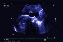
Presenter Nicole Adelson from Hofstra University in Hempstead, NY, shared results showing that AI-based quantitative ultrasound can accurately assess fetal lung maturity.
“I created an AI model using convolutional neural networks (CNNs) to characterize fetal ultrasound images as pre-term or term, and that is based on the heterogeneity index that we can derive from images,” Adelson told AuntMinnie.
Lung immaturity is a leading cause of high mortality rates in pre-term infants, with respiratory distress syndrome being a complication from this condition. Current assessment methods are invasive and have limited accuracy, Adelson said.
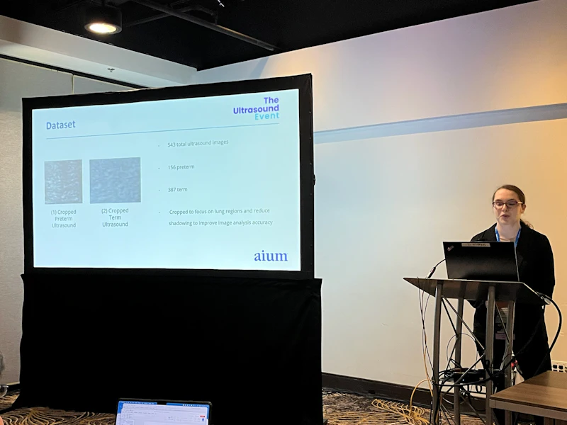 Nicole Adelson from Hofstra University presents findings at AIUM 2026 showing how an ultrasound-based AI model could help measure lung maturity in developing fetuses.AuntMinnie
Adelson and colleagues developed and tested their AI model to quantitatively analyze fetal lung ultrasound images by using dithering to highlight texture patterns. They also divided regions of interest into subregions for comparison and used a heterogeneity index to measure lung development. Higher heterogeneity means less air and immature lungs while higher homogeneity means more air and mature lungs.
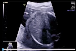
The team trained its model on 543 ultrasound images (156 pre-term, 387 term) and employed five-fold cross-validation for reliability.
The model achieved a validation accuracy of 92% and training accuracy of 88%. Adelson also reported stable loss throughout training and highlighted the model’s strong performance.
Nicole Adelson explains how her team's AI ultrasound approach could help with fetal lung assessment and shares her team's goal for the AI model.
Adelson told AuntMinnie the results are “very promising” and could help address fetal morbidity. She also said future improvements could include using Bayesian optimization to improve hyperparameters, expanding the team’s dataset with more images, and further using transfer learning.
The team’s goal is to create a portable ultrasound system that can allow for real-time clinical assessment.
“Essentially, we will have an app that will allow you to select the ultrasound image as well as the region of interest, and it will automatically return to you whether it’s pre-term or term as well as the heterogeneity index,” Adelson said. “This will improve maternal and fetal outcomes with physicians using this alongside their clinical practice and expand quantitative assessment to adult lung images.”
For more of AuntMinnie's coverage from AIUM 2026, click here.