PHILADELPHIA -- AI is helping sonographers all over the world deliver care to underserved areas and relieving some of the workload for imagers, according to a keynote presentation given May 28 at the American Institute of Ultrasound in Medicine (AIUM) annual meeting.
Modern video and multimodal deep learning methods can automatically analyze ultrasound video and support clinical decision-making by finding more information compared to conventional methods. This was one key takeaway from Dr. Alison Noble from the University of Oxford in the U.K., who presented her keynote on AI’s evolving role in ultrasound.
“It’s a very exciting time, because we’re starting to see some of the basic tools for measurements, for example, start to appear in systems,” Noble told AuntMinnie. “But also, in terms of things like ultrasound guidance, this is still very much in the research area. And I think in the future, we’re going to start to see some of the capabilities going to working systems and start to be available in the future.”
AI continues to show promise in medical imaging as an assistant for reading images and alleviating high-volume workloads for imagers. But even as the technology advances, some barriers to AI adoption persist. Noble said some of these include developing trust in using the technology and understanding what some of the use cases are.
“The technical capabilities are there, but sometimes when you try them out in the clinic, things might not work,” she said. “But you also learn from that, where the right place is for the technology.”
And education on best practices toward clinical AI use will also be needed, according to Noble.
Alison Noble talks about the human element of using AI in clinical practice, including how sonographers can work side by side with the technology.
Noble is the Technikos Professor of Biomedical Engineering in Oxford's Department of Engineering Science and is a professorial fellow of St Hilda’s College. She previously directed the Oxford Institute of Biomedical Engineering.
Her group's work focuses on bringing machine learning applications to medical ultrasound, as well as cardiac MRI and cell image analyses.
Current projects
Noble outlined several research projects that she and colleagues are leading, which are studying the use of AI in ultrasound education and clinical use in low- to middle-income countries.
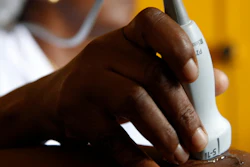
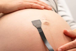
In Kenya, the group is exploring the capabilities of TraCer, a fully portable system uses a low-cost probe to capture fetal ultrasound videos. The system is integrated with a wireless ultrasound probe (Konted), with the application running on a standard Android tablet. Noble said this approach allows health workers to perform accurate pregnancy dating with minimal training and without costly equipment.
Another project is the Computer-Assisted Low-Cost Point-of-Care UltraSound (CALOPUS) Study being led in India. Noble and colleagues found in a 2022 study that this AI-assisted technique improved visualization of the cervix and classification of placental location.
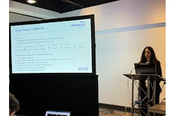
 Alison Noble, PhD, from the University of Oxford shares work that she and colleagues have been working on toward AI in ultrasound at AIUM 2026.
A study published in 2025 showed that AI aid in estimating gestational age led to reduced clinician mean absolute error (MAE) from 23.5 days to 15.7 days. And model explanations further reduced this to 14.3 days, with participants who found model explanations to be helpful showing improved performance. The researchers also found that while model explanations can improve confidence among sonographers, they can also reduce self-reported model trust.
“This is showing human attitude toward AI, but it’s showing that explanations do not always help,” Noble said. “It also relates to part of acceptable use is related to trust in using AI.”
Tracking models and emerging applications
AI may also help measure skills among sonographers. Noble talked about some measurement systems in ultrasound, including eye trackers for sonographer gazing, inertial measurement units for tracking probe movement, and audio for sonographer speech.
In one study published in 2021, Noble and colleagues put these into action and found they could assess cognitive workload from pupil diameter changes measured while ultrasound operators performed routine scans. The machine learning used in the study could discriminate between ultrasonographic tasks and scanning expertise.
Noble shares what she and colleagues are working on toward AI use in ultrasound.
Another study published in 2020 by Noble et al showed how an AI ultrasound model demonstrated high performance in predicting either probe movement toward the standard plane position or the next movement that an expert sonographer would perform. Researchers in 2022 also showed how a multimodal guidance approach from real-world ultrasound video signals, synchronized gaze, and probe motion could predict gaze movements and probe signals that an experienced sonographer would perform in routine obstetric scanning.
Finally, Noble shared two other emerging AI-based technologies for ultrasound, federated learning and human-AI collaboration. In federated learning, machine learning models can be trained on large-scale multi-source decentralized medical image data. And human-AI collaboration meanwhile could improve overall task performance beyond what humans and AI can do alone. Noble said this could also balance minimizing human workloads with maximizing trust in AI safety, accuracy, and efficiency.
Noble and colleagues led a study on how large language models (LLMs) can be used in a guided deferral system. Their pilot study focused on real-world spinal lumbar MRI text reports. All participants in the study showed improved accuracy with this human–AI collaboration approach compared to either LLMs or humans alone.
“It is becoming a truly interdisciplinary real-world data science field,” Noble said. “Originally, when I started in this space, it was engineers working with sonographers and clinicians. And now it’s bringing in human behavior scientists who experiment in psychology.”
For more of AuntMinnie's coverage from AIUM 2026, click here.