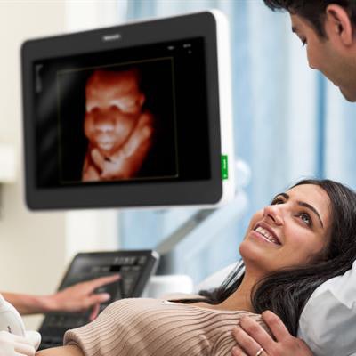
Philips Healthcare has launched a new premium ultrasound scanner family called Epiq Elite for general imaging and ob/gyn applications.
The scanner comes in two flavors: Epiq Elite for General Imaging is tailored to the specific needs of vascular assessment, while Epiq Elite for Obstetrics & Gynecology is designed to provide excellent image quality and lifelike 3D scans for better fetal assessment at all stages of pregnancy, according to the company.
The platform includes the company's Ultimate Ultrasound Solution for Vascular Assessment package, which gives users insight into the progression of vascular disease by combining 3D and 4D imaging. It also offers a simplified workflow and complementary clinical tools to monitor and assess vascular disease, Philips said.
Philips' TrueVue technology enables clinicians to manipulate a virtual light source around 3D fetal images to produce lifelike images. Image courtesy of Philips.Using the company's xMatrix transducers, users can acquire 3D vascular images that enable clinicians to easily visualize vessels directly and evaluate the spatial location and composition of plaque. The scanner also includes a live xPlane imaging mode that allows clinicians to acquire two planes simultaneously for better accuracy in data collection while reducing exam time by 20%.
Meanwhile, the Epiq Elite for Obstetrics & Gynecology scanner includes new transducers and software that improve fetal assessment and meet the specific needs of ob/gyn patients, especially during the early stages of pregnancy. For example, V9-2 is a lightweight, ergonomic transducer that is the first high-frequency transducer based on the company's PureWave technology. When used with Philips' TrueVue image processing mode, Epiq Elite allows clinicians to manipulate a virtual light source around 3D fetal images to produce incredibly lifelike results, according to the company.
In addition, Philips' aBiometry Assist application uses what Philips calls "anatomical intelligence" to automate the time sonographers spend gathering measurements. This reduces exam time and allows clinicians to spend more time with patients.
Philips plans to show Epiq Elite for Obstetrics & Gynecology at the upcoming Society of Maternal-Fetal Medicine (SMFM) conference in Las Vegas, and it will show Epiq Elite for General Imaging at ECR 2019 in Vienna.

![Examples of ultrasound findings and techniques. (A) Images in a 39-year-old male patient with a mass in the left thigh. The mass is heterogeneous on the B-mode US image (compared with the patient in D) and showed increased microvascularity (superb microvascular imaging [SMI]) and shear-wave elastography (SWE) values. Undifferentiated pleomorphic sarcoma was diagnosed at biopsy (with pleomorphic rhabdomyosarcoma in surgical specimen). (B) Images in an 18-year-old male patient with a mass in the left leg. The mass is hypoechoic on the B-mode image, with no other findings suggestive of malignancy. The lesion is in contact with the cortex of the tibia, which is slightly irregular. CT revealed a doubtful anteromedial tibial erosion. The microvascular study demonstrated high vascularization, suggestive of malignancy. Periosteal Ewing sarcoma was diagnosed with both histologic and immunohistochemical confirmation. (C) Images in a 69-year-old female patient with a lump growing on the outside of the left leg. Multiple SWE examinations were performed (please note the high values obtained in the measurements, whereas the color map highlights the stiffness relative to adjacent tissues). SMI showed areas of increased vascularization to target for sampling. Undifferentiated spindle cell sarcoma was diagnosed at biopsy, with residual leiomyosarcoma in the surgical specimen after neoadjuvant therapy. (D) Images in a 56-year-old female patient with a mass in the right thigh. The mass is heterogeneous at both B-mode ultrasound (similar to patient A) and MRI (coronal T2-weighted spectral attenuated inversion recovery [SPAIR]; T1-weighted pre-contrast and postcontrast imaging), which even shows uptake after the administration of paramagnetic contrast material, which is traditionally suggestive of malignancy. Low values at SMI and elastography are suggestive of benignity. Spindle cell lipoma was diagnosed at biopsy, with atypical spindle cell lipomatous tumor in the surgical specimen.](https://img.auntminnie.com/mindful/smg/workspaces/default/uploads/2026/08/images-radiol250278fig2.APCFLSvX6p.jpg?auto=format%2Ccompress&fit=crop&h=100&q=70&w=100)


![Examples of ultrasound findings and techniques. (A) Images in a 39-year-old male patient with a mass in the left thigh. The mass is heterogeneous on the B-mode US image (compared with the patient in D) and showed increased microvascularity (superb microvascular imaging [SMI]) and shear-wave elastography (SWE) values. Undifferentiated pleomorphic sarcoma was diagnosed at biopsy (with pleomorphic rhabdomyosarcoma in surgical specimen). (B) Images in an 18-year-old male patient with a mass in the left leg. The mass is hypoechoic on the B-mode image, with no other findings suggestive of malignancy. The lesion is in contact with the cortex of the tibia, which is slightly irregular. CT revealed a doubtful anteromedial tibial erosion. The microvascular study demonstrated high vascularization, suggestive of malignancy. Periosteal Ewing sarcoma was diagnosed with both histologic and immunohistochemical confirmation. (C) Images in a 69-year-old female patient with a lump growing on the outside of the left leg. Multiple SWE examinations were performed (please note the high values obtained in the measurements, whereas the color map highlights the stiffness relative to adjacent tissues). SMI showed areas of increased vascularization to target for sampling. Undifferentiated spindle cell sarcoma was diagnosed at biopsy, with residual leiomyosarcoma in the surgical specimen after neoadjuvant therapy. (D) Images in a 56-year-old female patient with a mass in the right thigh. The mass is heterogeneous at both B-mode ultrasound (similar to patient A) and MRI (coronal T2-weighted spectral attenuated inversion recovery [SPAIR]; T1-weighted pre-contrast and postcontrast imaging), which even shows uptake after the administration of paramagnetic contrast material, which is traditionally suggestive of malignancy. Low values at SMI and elastography are suggestive of benignity. Spindle cell lipoma was diagnosed at biopsy, with atypical spindle cell lipomatous tumor in the surgical specimen.](https://img.auntminnie.com/mindful/smg/workspaces/default/uploads/2026/08/images-radiol250278fig2.APCFLSvX6p.jpg?auto=format%2Ccompress&fit=crop&h=112&q=70&w=112)