PHILADELPHIA -- A multimodal approach featuring ultrasound shows how nanoplastics are retained in endometrial polyps, according to research presented May 28 at the American Institute of Ultrasound in Medicine (AIUM) annual meeting.
In her presentation, Hailie Ciomperlik, MD, from Baylor University in Waco, TX, showed that more nanoplastics are retained in larger polyps and that this accumulation varies by lesion type.
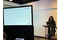
 Hailie Ciomperlik, MD, from Baylor University presents her team's results at AIUM 2026, showing how a multimodal approach featuring ultrasound shows nanoplastic retention within endometrial polyps.
“While causality cannot be inferred, the findings introduce an environmental dimension to endometrial polyp biology with potential implications for infertility,” she said.
Microplastics and nanoplastics have come under the spotlight in recent years as prior research shows how these are detected in human reproductive tissues. Experimental models suggest that nanoplastics change stromal proliferation and inflammation in endometrial polyps.
However, there is currently no data linking tissue polymer levels to specific polyp morphologic characteristics.
Ciomperlik and colleagues studied nanoplastic retention in the presence of multiple or larger polyps. Their retrospective study consisted of 28 women, including 13 with polyps, 13 being healthy controls, and two being saline controls.
The team used a multimodal approach consisting of ultrasound, saline infusion sonogram (SIS), hysteroscopy, and pathology.
Ciomperlik reported the following predominant micro- and nanoplastics found: polyvinyl chloride (PVC), polyethylene terephthalate (PET), and polymethyl methacrylate (PMMA). And the cumulative nanoplastic burden achieved a median of 897 micrograms per gram (µg/g).
Larger polyps (>10 mm) showed associations with higher odds per doubling of nanoplastic load. These included odds ratios (ORs) of 1.76 for PMMA, 1.48 for PET, and 1.18 for PVC. Cumulative burden also showed a positive association with polyp size (OR = 1.21). However, none of these associations reached statistical significance, Ciomperlik said.
Finally, Ciomperlik reported a consistent direction with these findings. Nanoplastic load was about 1.2 to 1.8 times greater in larger polyps versus smaller lesions, with the fundus having the largest burden and the posterior wall having the least burden.
Ciomperlik said this suggests spatial heterogeneity in uterine polymer retention and is possibly related to uterine fluid dynamics, vascularity, or tissue architecture.
“Findings were consistent across hysteroscopic and pathology-based models,” she said. “The results suggest polymer-specific environmental exposure may contribute to polyp biology.
Ciomperlik said the team’s next steps include mechanistic studies and larger cohorts to define the biological effects of nanoplastics on human reproductive systems.
For more of AuntMinnie's coverage from AIUM 2026, click here.