Dear AuntMinnie Member,
Proton MR spectroscopy has always been a powerful tool for assessing metabolic changes in the body. But what if you performed the technique with a 7-tesla MRI scanner?
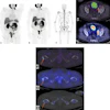
That's what researchers in Austria wanted to find out. They used a 7-tesla MRI scanner to perform proton spectroscopy in an effort to identify substances produced by a person's metabolism that could indicate the presence of multiple sclerosis. Find out how well it worked in an article in our MRI Community.
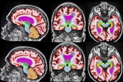
While you're in the community, check out our article on the best sequences to use as part of an automated MR volumetry technique for brain imaging. And don't miss a story from earlier in the week on whether women who are eligible for supplemental breast MRI screening because of their higher risk of breast cancer are actually getting the scans.
All these stories are available in our MRI Community.
Ultrasound for dense breasts
Ultrasound has emerged as a valuable tool for screening women who may have dense breast tissue. The modality's role should get further clarified with the release soon in the U.S. of national standards for when it should be used as an adjunct to mammography in women with dense breasts.
We're pleased to announce a new special article on breast ultrasound in our Ultrasound Community. Contributing writer Emily Hayes talked to key opinion leaders in breast ultrasound to get their take on how and when it should be used in women with dense breast tissue.
In other important ultrasound news, diagnostic ultrasound proved superior to other methods when it came to assessing margins of cancers being excised during breast-conserving surgery, according to researchers from Denmark.
And in a study out of Johns Hopkins University, researchers suggested that diagnostic breast ultrasound scans to assess suspicious masses found on mammography screening should be postponed if the woman recently received a COVID-19 vaccine.
January 12 webinar on AI
Finally, our free webinar on article intelligence (AI) in radiology is coming up on January 12. Have you reserved your seat yet? We have a great lineup of key opinion leaders, including Dr. Erik Ranschaert, PhD; Dr. Geraldine McGinty; and Wiro Niessen, PhD. Register now to participate in this important event.