Photon-counting CT (PCCT) performs comparably to MRI for characterizing breast lesions, detecting additional lesions, and assessing lymph node involvement, making it a potential alternative for supplemental breast imaging in cancer evaluation.
- PCCT achieved similar sensitivity to MRI for detecting additional breast lesions, with only a 7% difference in performance.
- PCCT demonstrated higher accuracy than MRI for assessing lymph node metastasis, with a 10% sensitivity advantage.
- The study included 126 women who underwent both PCCT and MRI imaging for comparison of diagnostic capabilities.
- PCCT showed good to excellent agreement with MRI for lesion characterization and tumor staging (κ = 0.57-0.96).
- PCCT may enable single-session pre-treatment breast cancer evaluation, potentially improving breast-conserving surgery rates.
Photon-counting CT (PCCT) could be a suitable alternative to MRI for supplemental breast imaging, suggest findings published September 1 in Radiology.
PCCT was on par with MRI for characterizing breast lesions and finding additional lesions, wrote a research group led by Qiuyi Fu, MD, from Ruijin Hospital and Shanghai Jiao Tong University in China. And while PCCT had limited success in finding breast microcalcifcations, it achieved high nodal accuracy similar to that of MRI.
“PCCT may serve as an integrated, single-session modality for pre-treatment breast cancer evaluation,” the Fu group wrote.
Since its clearance by the U.S. Food and Drug Administration (FDA) in 2021, PCCT has shown its potential in improving diagnostic imaging. Proponents highlight PCCT’s better spatial resolution, contrast to noise ratio, and dose efficiency. However, its clinical utility for breast cancer is not completely defined, the researchers noted.
Fu and colleagues studied PCCT’s feasibility for pre-treatment breast cancer assessment. They compared the modality’s performance with MRI, full-field digital mammography, and fluorine 18 (18F) fluorodeoxyglucose (FDG) PET/CT.
The study included 126 women with an average age of 58.1 years who had BI-RADS 4C or higher lesions at ultrasound or full-field digital mammography. The women underwent either supplemental MRI and multiphasic contrast-enhanced PCCT. 18F-FDG PET/CT was performed in a subset with locally advanced disease.
Four radiologists independently evaluated lesion morphologic characteristics, additional findings, and clinical staging. The radiologists achieved good to excellent interreader agreement across PCCT, MRI, and full-field digital mammography.
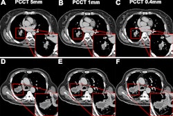
 Detection and extent of BI-RADS suspicious calcifications (amorphous, coarse heterogeneous, fine pleomorphic, and fine linear or fine linear branching) at axial photon-counting CT (PCCT) versus full-field digital mammography (first three images, craniocaudal view; last image, mediolateral oblique view). Calcifications found on mammography showed only partial visualization at ultrahigh-resolution PCCT (0.2 mm), while amorphous calcifications could not be distinguished from image noise. Corresponding abnormal enhancement was clearly depicted on contrast-enhanced PCCT images.RSNA
PCCT agreed with MRI for lesion characterization (κ = 0.57-0.96) and clinical T categorization (κ = 0.86-0.88). The team noted the highest agreement with pathologic size between the two modalities (intraclass correlation coefficient, 0.70-0.81).
For 46 pathologically confirmed additional lesions, PCCT achieved higher sensitivity than full-field digital mammography (difference, 44%) and similar sensitivity compared to MRI (difference, 7%). PCCT however missed 44% of microcalcifications found on full-field digital mammography.
PCCT achieved higher sensitivity and accuracy than MRI for assessing pathologically confirmed nodal metastasis, with differences of 10% and 6%, respectively.
And for clinical N category cancers, PCCT agreed with PET/CT (κ = 0.82; n = 19). Two distant metastases found on PCCT were consistent with 18F-FDG PET/CT and pathologic findings.
The study authors called for future research to validate breast PCCT in multicenter studies. This includes developing PCCT-specific BI-RADS descriptors, improving ultralow-dose protocols, determining the best acquisition position via prone versus supine comparisons, and examining the impact of clinical and economic outcomes in larger cohorts.
“If PCCT more accurately reflects the extent of the pathologic tumor, unnecessary tissue removal may be reduced and breast-conserving surgery rates, which are currently reduced by MRI-based staging results, could be improved,” the authors wrote.
Read the full study here.