Point-of-care ultrasound (POCUS) in emergency departments significantly reduces patient wait times and time to diagnosis in early pregnancy cases by approximately 60 and 81 minutes respectively, while maintaining the same safety profile and ectopic pregnancy detection rates as standard ultrasound.
- Time savings: POCUS reduced emergency department length of stay by 59.53 minutes and time to definitive diagnosis by 80.93 minutes compared to standard ultrasound
- Safety maintained: No statistically significant difference in missed ectopic pregnancy cases between POCUS and standard ultrasound approaches
- Study scope: Analysis of 13 studies involving 4,057 early pregnancy patients (1,848 with POCUS, 2,209 with standard ultrasound)
- Clinical impact: POCUS enables rapid identification of intrauterine pregnancy and accelerates bedside decision-making in emergency settings
- Research call: Experts recommend high-quality, multicenter randomized trials to further validate POCUS effectiveness and generalizability
Point-of-care ultrasound (POCUS) can improve emergency pregnancy care, according to findings published August 30 in the Journal of Emergency Medicine.
POCUS in the emergency department led to reduced length of stay and time to definitive diagnosis while not leading to more missed diagnoses of ectopic pregnancy, wrote a team led by Moaz Safwan, MD, from Sulaiman Al Rajhi University in Saudi Arabia.
“In this context, emergency department-performed POCUS has emerged as a pivotal diagnostic tool to provide a rapid identification of intrauterine pregnancy and accelerate decision-making at the bedside,” the Al Rajhi team wrote.
Ectopic pregnancy is a leading cause of pregnancy-related maternal mortality, making timely first-trimester evaluation of pregnant women critical. Women present to the emergency department with non-specific symptoms such as abdominal pain and vaginal bleeding, symptoms that can overlap with other diseases and present diagnostic challenges.
Transvaginal or transabdominal ultrasound are performed for early pregnancy evaluation. But logistical constraints can limit access to these exams, such as during peak demand or after hours. Previous research suggests that POCUS can help reduce these burdens by decreasing length of stay without compromising care.
Safwan and colleagues compared the effectiveness of POCUS with that of standard radiology or obstetric ultrasound for early pregnancy assessment in acute care settings. They focused on differences between these approaches for the following measures: emergency department length of stay, time to diagnosis, time to surgery for patients with ectopic pregnancy, and rate of missed ectopic cases.
The team reviewed 13 studies from four databases with 4,057 early pregnancy patients. These included 1,848 who underwent POCUS and 2,209 who underwent standard ultrasound.
POCUS in the emergency department led to statistically significant reductions in length of stay and time to definitive diagnosis. These included weighted mean differences of -59.53 minutes (p < 0.00001) and -80.93 minutes (p < 0.0001), respectively.
And pooled analysis showed no statistically significant difference in the rate of missed ectopic cases between POCUS and standard ultrasound (risk ratio, 1.22; p = 0.73).
The researchers noted that their results should be interpreted with caution due to study heterogeneity, observational designs, and potential publication bias.
They called for high-quality, multicenter randomized studies to “better define the diagnostic accuracy, safety, and generalizability” of emergency department POCUS in early pregnancy evaluation.
Read the full study here.

![Examples of ultrasound findings and techniques. (A) Images in a 39-year-old male patient with a mass in the left thigh. The mass is heterogeneous on the B-mode US image (compared with the patient in D) and showed increased microvascularity (superb microvascular imaging [SMI]) and shear-wave elastography (SWE) values. Undifferentiated pleomorphic sarcoma was diagnosed at biopsy (with pleomorphic rhabdomyosarcoma in surgical specimen). (B) Images in an 18-year-old male patient with a mass in the left leg. The mass is hypoechoic on the B-mode image, with no other findings suggestive of malignancy. The lesion is in contact with the cortex of the tibia, which is slightly irregular. CT revealed a doubtful anteromedial tibial erosion. The microvascular study demonstrated high vascularization, suggestive of malignancy. Periosteal Ewing sarcoma was diagnosed with both histologic and immunohistochemical confirmation. (C) Images in a 69-year-old female patient with a lump growing on the outside of the left leg. Multiple SWE examinations were performed (please note the high values obtained in the measurements, whereas the color map highlights the stiffness relative to adjacent tissues). SMI showed areas of increased vascularization to target for sampling. Undifferentiated spindle cell sarcoma was diagnosed at biopsy, with residual leiomyosarcoma in the surgical specimen after neoadjuvant therapy. (D) Images in a 56-year-old female patient with a mass in the right thigh. The mass is heterogeneous at both B-mode ultrasound (similar to patient A) and MRI (coronal T2-weighted spectral attenuated inversion recovery [SPAIR]; T1-weighted pre-contrast and postcontrast imaging), which even shows uptake after the administration of paramagnetic contrast material, which is traditionally suggestive of malignancy. Low values at SMI and elastography are suggestive of benignity. Spindle cell lipoma was diagnosed at biopsy, with atypical spindle cell lipomatous tumor in the surgical specimen.](https://img.auntminnie.com/mindful/smg/workspaces/default/uploads/2026/08/images-radiol250278fig2.APCFLSvX6p.jpg?auto=format%2Ccompress&fit=crop&h=100&q=70&w=100)


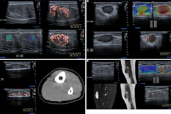
![Examples of ultrasound findings and techniques. (A) Images in a 39-year-old male patient with a mass in the left thigh. The mass is heterogeneous on the B-mode US image (compared with the patient in D) and showed increased microvascularity (superb microvascular imaging [SMI]) and shear-wave elastography (SWE) values. Undifferentiated pleomorphic sarcoma was diagnosed at biopsy (with pleomorphic rhabdomyosarcoma in surgical specimen). (B) Images in an 18-year-old male patient with a mass in the left leg. The mass is hypoechoic on the B-mode image, with no other findings suggestive of malignancy. The lesion is in contact with the cortex of the tibia, which is slightly irregular. CT revealed a doubtful anteromedial tibial erosion. The microvascular study demonstrated high vascularization, suggestive of malignancy. Periosteal Ewing sarcoma was diagnosed with both histologic and immunohistochemical confirmation. (C) Images in a 69-year-old female patient with a lump growing on the outside of the left leg. Multiple SWE examinations were performed (please note the high values obtained in the measurements, whereas the color map highlights the stiffness relative to adjacent tissues). SMI showed areas of increased vascularization to target for sampling. Undifferentiated spindle cell sarcoma was diagnosed at biopsy, with residual leiomyosarcoma in the surgical specimen after neoadjuvant therapy. (D) Images in a 56-year-old female patient with a mass in the right thigh. The mass is heterogeneous at both B-mode ultrasound (similar to patient A) and MRI (coronal T2-weighted spectral attenuated inversion recovery [SPAIR]; T1-weighted pre-contrast and postcontrast imaging), which even shows uptake after the administration of paramagnetic contrast material, which is traditionally suggestive of malignancy. Low values at SMI and elastography are suggestive of benignity. Spindle cell lipoma was diagnosed at biopsy, with atypical spindle cell lipomatous tumor in the surgical specimen.](https://img.auntminnie.com/mindful/smg/workspaces/default/uploads/2026/08/images-radiol250278fig2.APCFLSvX6p.jpg?auto=format%2Ccompress&fit=crop&h=167&q=70&w=250)
![Examples of ultrasound findings and techniques. (A) Images in a 39-year-old male patient with a mass in the left thigh. The mass is heterogeneous on the B-mode US image (compared with the patient in D) and showed increased microvascularity (superb microvascular imaging [SMI]) and shear-wave elastography (SWE) values. Undifferentiated pleomorphic sarcoma was diagnosed at biopsy (with pleomorphic rhabdomyosarcoma in surgical specimen). (B) Images in an 18-year-old male patient with a mass in the left leg. The mass is hypoechoic on the B-mode image, with no other findings suggestive of malignancy. The lesion is in contact with the cortex of the tibia, which is slightly irregular. CT revealed a doubtful anteromedial tibial erosion. The microvascular study demonstrated high vascularization, suggestive of malignancy. Periosteal Ewing sarcoma was diagnosed with both histologic and immunohistochemical confirmation. (C) Images in a 69-year-old female patient with a lump growing on the outside of the left leg. Multiple SWE examinations were performed (please note the high values obtained in the measurements, whereas the color map highlights the stiffness relative to adjacent tissues). SMI showed areas of increased vascularization to target for sampling. Undifferentiated spindle cell sarcoma was diagnosed at biopsy, with residual leiomyosarcoma in the surgical specimen after neoadjuvant therapy. (D) Images in a 56-year-old female patient with a mass in the right thigh. The mass is heterogeneous at both B-mode ultrasound (similar to patient A) and MRI (coronal T2-weighted spectral attenuated inversion recovery [SPAIR]; T1-weighted pre-contrast and postcontrast imaging), which even shows uptake after the administration of paramagnetic contrast material, which is traditionally suggestive of malignancy. Low values at SMI and elastography are suggestive of benignity. Spindle cell lipoma was diagnosed at biopsy, with atypical spindle cell lipomatous tumor in the surgical specimen.](https://img.auntminnie.com/mindful/smg/workspaces/default/uploads/2026/08/images-radiol250278fig2.APCFLSvX6p.jpg?auto=format%2Ccompress&fit=crop&h=112&q=70&w=112)