An artificial intelligence (AI) model appears effective for detecting malpositioned endotracheal tubes on patient x-rays, according to a study presented at the American Roentgen Ray Society (ARRS) annual meeting.
A group led by researchers at Seoul National University Hospital and including colleagues at AI software firm Lunit tested a deep-learning model trained on chest x-rays from intubated patients in intensive care units. The model performed well and could potentially be used to alert clinicians of cases where tubes need to be repositioned, according to the group.
"A deep-learning system exhibited excellent performance in identifying the presence of ET [endotracheal tube] and malposition of ET on chest radiographs," noted corresponding author Dr. Eui Jin Hwang, in a thoracic imaging poster presentation.
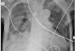
Chest x-rays are used to evaluate tube placements after clinicians have intubated patients, as improperly positioned tubes can cause complications. Tubes inserted in patients too deeply can cause contralateral lung collapse or ipsilateral hyperinflation, while shallowly inserted tubes can result in air leaks or vocal cord injuries, the authors explained. They hypothesized that a deep-learning AI model could help evaluate tube placements and ultimately reduce morbidity rates associated with these complications.
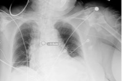
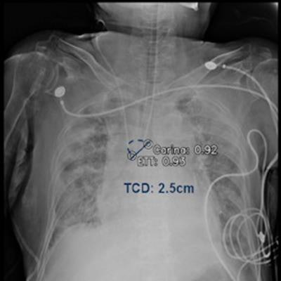
The team developed a model based on 539 consecutive portable chest x-rays taken immediately after tube insertions in patients in internal care units at their hospital. The model was trained to identify the presence versus absence of endotracheal tubes and correct placements based on distances between the tube tip and the ridge at the end of the windpipe, or the tracheal carina.
A slide presented by researchers at the ARRS meeting in Hawaii showed measurements used by an AI model for detecting correct endotracheal tube placements in x-rays of intensive care unit (ICU) patients. Image courtesy of Dr. Eui Jin Hwang.Tubes were malpositioned in 76 of the chest x-rays. In a comparison with an experienced thoracic radiologist and one trainee who independently evaluated the placements, the model achieved similar accuracy, the group found.
For deep malpositions of tubes, the model achieved an area under the receiver operating characteristic curve (AUC) of 0.96, and for shallow malpositions of tubes, the model achieved an AUC of 0.97, the researchers reported.
"A DL system showed excellent interrater agreement compared with radiologists in measuring [distance between the tube tip and carina] on chest radiographs," the authors wrote.
With further development and validation, the AI model could facilitate early notification of malpositioned tubes to clinicians for repositioning, they concluded.


![Examples of ultrasound findings and techniques. (A) Images in a 39-year-old male patient with a mass in the left thigh. The mass is heterogeneous on the B-mode US image (compared with the patient in D) and showed increased microvascularity (superb microvascular imaging [SMI]) and shear-wave elastography (SWE) values. Undifferentiated pleomorphic sarcoma was diagnosed at biopsy (with pleomorphic rhabdomyosarcoma in surgical specimen). (B) Images in an 18-year-old male patient with a mass in the left leg. The mass is hypoechoic on the B-mode image, with no other findings suggestive of malignancy. The lesion is in contact with the cortex of the tibia, which is slightly irregular. CT revealed a doubtful anteromedial tibial erosion. The microvascular study demonstrated high vascularization, suggestive of malignancy. Periosteal Ewing sarcoma was diagnosed with both histologic and immunohistochemical confirmation. (C) Images in a 69-year-old female patient with a lump growing on the outside of the left leg. Multiple SWE examinations were performed (please note the high values obtained in the measurements, whereas the color map highlights the stiffness relative to adjacent tissues). SMI showed areas of increased vascularization to target for sampling. Undifferentiated spindle cell sarcoma was diagnosed at biopsy, with residual leiomyosarcoma in the surgical specimen after neoadjuvant therapy. (D) Images in a 56-year-old female patient with a mass in the right thigh. The mass is heterogeneous at both B-mode ultrasound (similar to patient A) and MRI (coronal T2-weighted spectral attenuated inversion recovery [SPAIR]; T1-weighted pre-contrast and postcontrast imaging), which even shows uptake after the administration of paramagnetic contrast material, which is traditionally suggestive of malignancy. Low values at SMI and elastography are suggestive of benignity. Spindle cell lipoma was diagnosed at biopsy, with atypical spindle cell lipomatous tumor in the surgical specimen.](https://img.auntminnie.com/mindful/smg/workspaces/default/uploads/2026/08/images-radiol250278fig2.APCFLSvX6p.jpg?auto=format%2Ccompress&fit=crop&h=100&q=70&w=100)


![Examples of ultrasound findings and techniques. (A) Images in a 39-year-old male patient with a mass in the left thigh. The mass is heterogeneous on the B-mode US image (compared with the patient in D) and showed increased microvascularity (superb microvascular imaging [SMI]) and shear-wave elastography (SWE) values. Undifferentiated pleomorphic sarcoma was diagnosed at biopsy (with pleomorphic rhabdomyosarcoma in surgical specimen). (B) Images in an 18-year-old male patient with a mass in the left leg. The mass is hypoechoic on the B-mode image, with no other findings suggestive of malignancy. The lesion is in contact with the cortex of the tibia, which is slightly irregular. CT revealed a doubtful anteromedial tibial erosion. The microvascular study demonstrated high vascularization, suggestive of malignancy. Periosteal Ewing sarcoma was diagnosed with both histologic and immunohistochemical confirmation. (C) Images in a 69-year-old female patient with a lump growing on the outside of the left leg. Multiple SWE examinations were performed (please note the high values obtained in the measurements, whereas the color map highlights the stiffness relative to adjacent tissues). SMI showed areas of increased vascularization to target for sampling. Undifferentiated spindle cell sarcoma was diagnosed at biopsy, with residual leiomyosarcoma in the surgical specimen after neoadjuvant therapy. (D) Images in a 56-year-old female patient with a mass in the right thigh. The mass is heterogeneous at both B-mode ultrasound (similar to patient A) and MRI (coronal T2-weighted spectral attenuated inversion recovery [SPAIR]; T1-weighted pre-contrast and postcontrast imaging), which even shows uptake after the administration of paramagnetic contrast material, which is traditionally suggestive of malignancy. Low values at SMI and elastography are suggestive of benignity. Spindle cell lipoma was diagnosed at biopsy, with atypical spindle cell lipomatous tumor in the surgical specimen.](https://img.auntminnie.com/mindful/smg/workspaces/default/uploads/2026/08/images-radiol250278fig2.APCFLSvX6p.jpg?auto=format%2Ccompress&fit=crop&h=112&q=70&w=112)