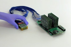
Engineers at the University of California San Diego have developed a soft, wearable ultrasound patch capable of continuously monitoring a fetus and umbilical cord for hours at a time, according to a study published May 26 in Nature Biotechnology and highlighted by UC San Diego Today.
The research was conducted by a team led by co-first author Geonho (Tom) Park, a chemical and nano engineering PhD candidate at the UC San Diego Jacobs School of Engineering.
The patch uses autonomous tracking algorithms to follow the umbilical cord as it moves, enabling consistent measurements as the mother or fetus changes position, the researchers said. In a collaboration study between UC San Diego Health and John Radcliffe Hospital at the University of Oxford in England, the device produced measurements comparable to standard handheld ultrasound across 62 pregnancies, including those complicated by gestational diabetes, pre-eclampsia, hypertension, and abnormal fetal growth.
A wearable ultrasound patch could provide continuous monitoring of the fetus and umbilical cord during pregnancy.Geonho (Tom) Park and UC San Diego
In one case, the patch detected prolonged abnormal fetal signals that prompted an early cesarean delivery at 29 weeks, which the researchers said may have helped save the baby's life.
The team plans to integrate the patch into a compact wireless electronic system for future use.
Access the full study here and UC San Diego Today's report here.








![Examples of ultrasound findings and techniques. (A) Images in a 39-year-old male patient with a mass in the left thigh. The mass is heterogeneous on the B-mode US image (compared with the patient in D) and showed increased microvascularity (superb microvascular imaging [SMI]) and shear-wave elastography (SWE) values. Undifferentiated pleomorphic sarcoma was diagnosed at biopsy (with pleomorphic rhabdomyosarcoma in surgical specimen). (B) Images in an 18-year-old male patient with a mass in the left leg. The mass is hypoechoic on the B-mode image, with no other findings suggestive of malignancy. The lesion is in contact with the cortex of the tibia, which is slightly irregular. CT revealed a doubtful anteromedial tibial erosion. The microvascular study demonstrated high vascularization, suggestive of malignancy. Periosteal Ewing sarcoma was diagnosed with both histologic and immunohistochemical confirmation. (C) Images in a 69-year-old female patient with a lump growing on the outside of the left leg. Multiple SWE examinations were performed (please note the high values obtained in the measurements, whereas the color map highlights the stiffness relative to adjacent tissues). SMI showed areas of increased vascularization to target for sampling. Undifferentiated spindle cell sarcoma was diagnosed at biopsy, with residual leiomyosarcoma in the surgical specimen after neoadjuvant therapy. (D) Images in a 56-year-old female patient with a mass in the right thigh. The mass is heterogeneous at both B-mode ultrasound (similar to patient A) and MRI (coronal T2-weighted spectral attenuated inversion recovery [SPAIR]; T1-weighted pre-contrast and postcontrast imaging), which even shows uptake after the administration of paramagnetic contrast material, which is traditionally suggestive of malignancy. Low values at SMI and elastography are suggestive of benignity. Spindle cell lipoma was diagnosed at biopsy, with atypical spindle cell lipomatous tumor in the surgical specimen.](https://img.auntminnie.com/mindful/smg/workspaces/default/uploads/2026/08/images-radiol250278fig2.APCFLSvX6p.jpg?auto=format%2Ccompress&fit=crop&h=112&q=70&w=112)


