High-resolution specimen PET/CT could help address positive margins in women undergoing surgery for breast cancer, suggest findings published June 17 in JAMA Surgery.
Specimen PET/CT imaging provided reliable intraoperative visualization in invasive ductal carcinoma and outperformed routine intraoperative margin assessment methods, wrote a team led by Menekse Göker, MD, from Cancer Research Institute Ghent in Belgium.
“The use of this integrated approach might lead to a substantial reduction of re-excision rates after breast-conserving surgery,” the Göker team wrote.
Positive margins occur in 12% to 30% of breast-conserving surgeries. These are tied to increased local recurrence risks, making accurate and efficient intraoperative margin assessment important.
Other imaging methods have been explored in this area but may disrupt workflows and may not be cost-effective depending on the modality. And while pathology analysis is accurate, its use is limited by longer turnaround times, labor intensiveness, and costs.
Göker and colleagues evaluated the success rate of intraoperative specimen PET/CT in addressing positive margins in women with invasive breast cancer.
The interventional nonrandomized clinical trial recruited 148 women from 2022 to 2025. Patients were followed up two weeks after surgery. Six European breast cancer centers took part in the study and integrated specimen PET/CT imaging into their routine surgical flows.
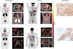
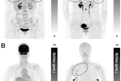
The women received an intravenous injection of low-dose F-18 FDG. The researchers analyzed PET/CT images of tumor specimens that were scanned intraoperatively. The preoperative tumor sizes ranged between 12 and 22 mm, with a median of 17 mm.
For the invasive component of invasive ductal carcinoma, success rates increased from 83.3% without intraoperative margin assessment to 86.9% with routine margin assessment. This further increased to 95.2% with specimen PET/CT (p < 0.001 vs. no margin assessment).
Across all study groups, success rates improved from 76.4% without margin assessment to 81.8% with routine margin assessment techniques. PET/CT further improved this to 91.9% (p < 0.001 vs. no margin assessment; p = 0.009 vs. standard-of-care margin assessment).
Along with lower rates of re-excision, the study authors highlighted PET/CT’s workflow advantages compared to margin assessment methods that could put a strain on resources.
“Overall, specimen PET-CT appears to balance sensitivity and specificity very well while offering the added advantage over other [margin assessment] modalities of 3D whole-specimen evaluation and easily fitting within the surgical workflow,” the authors wrote.
However, they also noted that PET/CT’s current implementation “may be limited by cost, availability, and technical requirements, potentially restricting its use to specialized centers.”
Read the full study here.