Contrast-enhanced mammography (CEM) maintains high performance in breast cancer screening and shows better specificity and accuracy during follow-up incidence screens compared to baseline screening in women at higher risk, offering a cost-effective alternative to MRI.
- Incidence CEM screening achieved 91.5% specificity and 91.4% accuracy, significantly higher than baseline screening's 83.4% specificity and 83.5% accuracy
- Study analyzed 6,911 CEM screens in 2,756 women from 2015 to 2021, comparing 1,575 baseline and 3,336 incidence screens
- Cancer detection rate remained consistent between baseline (19 per 1,000) and incidence (11.6 per 1,000) screens with no significant difference
Contrast-enhanced mammography (CEM) may have utility in screening women at higher risk of breast cancer, according to research published July 28 in Radiology.
Incidence screening with CEM maintains high performance and may offer more benefits over baseline screening, wrote a team of researchers led by Carol Lee, MD, from the Memorial Sloan Kettering Cancer Center in New York.
“The ability of contrast-enhanced mammography to help detect cancer during prevalence screening in women at increased risk for breast cancer is maintained in subsequent incidence screens, with better specificity and accuracy,” the Lee team wrote.
Despite the off-label indications for CEM, radiology researchers continue to explore the modality’s use in screening settings. Supporters have highlighted advantages of CEM over other breast imaging techniques, including higher sensitivity than ultrasound, digital mammography, and digital breast tomosynthesis (DBT). CEM also has a shorter study time and lower overall cost than breast MRI, with previous research suggesting that CEM's performance is non-inferior to MRI.
Lee and colleagues previously studied first-round screening with CEM in women at intermediate risk for breast cancer. For the current study, the researchers compared the performance of CEM in baseline versus incidence screens in women at higher risk.
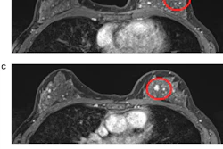
 Images in a 70-year-old asymptomatic woman with a prior excisional biopsy in the left breast yielding lobular carcinoma in situ 3 years earlier. Screening contrast-enhanced mammography (CEM) was performed, which was considered a prevalence screen as the woman had no prior breast MRI in the past 3 years and no prior CEM. (A) Low-energy (26–30 kVp) mediolateral oblique (MLO) and (B) craniocaudal (CC) views show surgical clips but no abnormality. (C) Recombined MLO and (D) CC images show an enhancing mass in the medial left breast (arrows). Biopsy revealed invasive lobular carcinoma. Size was 0.2 cm; tumor grade II/III; negative lymph nodes; and estrogen receptor–positive, progesterone receptor–positive, and human epidermal growth factor receptor 2–negative.RSNA
The retrospective study included 6,911 CEM screens in 2,756 women from 2015 to 2021. Of the total screens, 1,575 were baseline while 3,336 were incidence. The researchers categorized screens as baseline if there was no prior breast MRI in the past three years or no prior CEM.
Adjusting for the number of screens per individual, the team reported no significant difference in cancer detection rate between both screens. However, incidence CEM had higher specificity and accuracy than baseline CEM.
Comparison of CEM performance during baseline, incidence screening | |||
Measure | Baseline | Incidence | P value |
Cancer detection rate (per 1,000 screens) | 19 | 11.6 | 0.65 |
Incremental cancer detection rate (per 1,000 screens) | 11.4 | 6.7 | 0.1 |
Sensitivity | 87.1% | 86.1% | 0.6 |
Specificity | 83.4% | 91.5% | 0.008 |
Accuracy | 83.5% | 91.4% | 0.01 |
Contrast enhancement alone helped detect 18 of 30 cancers during baseline screening and 36 of 62 during incidence screening.
Finally, the team reported 14 interval cancers among the total exams. It found no evidence of a difference in cancers between the two groups (p = 0.4)
The study authors highlighted CEM as a “reasonable tool” for women with dense breasts, adding that it can also address health disparities for women who face barriers to MRI access.
“In addition, women have reported a preference for CEM over MRI,” they wrote.
The team also noted that a future article will provide complete details on the interval cancers found in this study.
While challenges remain in widespread adoption of CEM, the modality may “become a practical component” of breast cancer screening strategies for selected populations, according to an accompanying editorial by Vivianne Aguilera Freitas, MD, PhD, from the University of Toronto in Canada.
“Whether this potential ultimately translates into widespread implementation will depend on future studies evaluating not only diagnostic performance, but also patient outcomes, health care utilization, and real-world feasibility across diverse practice settings,” Freitas wrote.
Read the full study here.