Contrast-enhanced mammography (CEM) is on par with MRI in breast cancer screening for asymptomatic women, according to research published November 14 in Radiology.
Along with demonstrating CEM’s noninferiority to breast MRI, a team led by Jordana Phillips, MD, from the Beth Israel Deaconess Medical Center in Boston also found that CEM has a slight advantage over abbreviated MRI in this area, as well as superior performance to that of digital mammography.
“This study serves as a key step in moving toward an MRI alternative that can contribute to high-quality cancer care,” Phillips and colleagues wrote.
Previous studies have demonstrated the clinical utility of CEM in breast cancer screening as a potential alternative to MRI. However, the Phillips team noted a lack of data comparing the respective performance of the modalities to each other.
The researchers investigated whether CEM is comparable to standard breast MRI and abbreviated breast MRI for screening asymptomatic women, as well as whether it is superior to digital mammography.
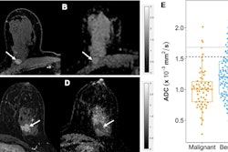
 A 47-year-old female participant with a suspicious mass noted at screening tomosynthesis and subsequent diagnostic ultrasound was recruited for contrast-enhanced mammography (CEM) and MRI. (A) No abnormality was noted on the conventional 2D mammogram showing dense breast tissue. A mass (arrows) was clearly observed on (B) the recombined CEM image and (C) the MRI scan. Both images show marked background parenchymal enhancement. Biopsy confirmed a 1.1-cm grade 2 invasive ductal carcinoma (estrogen receptor- and progesterone receptor-positive, human epidermal growth factor receptor 2-negative). Image courtesy of Radiology.
The team’s enriched reader study used imaging data prospectively collected from asymptomatic women at a single institution from 2014 to 2020, with 12 radiologists interpreting the images.
For CEM interpretation, the radiologists were first shown low-energy images as a surrogate for digital mammography. From there, they provided a forced BI-RADS score for up to three abnormalities, with the highest score being used as the case score. After that, they reviewed the full CEM exam and scored it similarly. After a minimum one-month washout, the radiologists interpreted abbreviated breast MRI and full MRI exams.
The researchers included data from 132 case sets. Of these, 74 were benign, 44 were malignant, and 14 were negative. The researchers found that CEM was noninferior to standard breast MRI and that digital mammography had inferior performance compared to the other tested modalities.
| Diagnostic performance of breast cancer screening modalities | |
|---|---|
| Modality | Area under the curve (AUC) |
| CEM | 0.91 |
| MRI | 0.91 |
| Abbreviated breast MRI | 0.89 |
| Digital mammography | 0.79 |
| *CEM showed statistical significance compared to digital mammography (p < 0.001). No significant differences were found between CEM, MRI, and abbreviated breast MRI. | |
On subset analysis, the team reported that CEM had significantly higher performance than that of digital mammography for examining both dense and nondense breast tissue (p = 0.02 and p < 0.001, respectively).
Additionally, CEM was on par with MRI and abbreviated breast MRI in diagnostic performance for both dense and non-dense tissue cases. CEM had AUC values of 0.89 and 0.94 for dense and nondense tissue, compared with AUC values of 0.89 and 0.95 for standard MRI, respectively. Abbreviated MRI meanwhile had AUC values of 0.87 and 0.94 for dense and nondense breast tissue, respectively.
The study authors highlighted that this study provides “important preliminary information for evaluating CEM as a viable screening option.” They also suggested that CEM’s performance may have been higher had digital imaging been used rather than the low-energy images of the same CEM exam as done in the study.
The full study can be found here.

![A normal mammogram confirmed by three-year radiologic follow-up illustrates reader-marked regions of interest (ROIs) during (A) unaided (round 1) and (B) artificial intelligence (AI)–assisted (round 2) reading. Each colored dot represents an ROI for recall by a human reader. Readers could mark more than one ROI per case, represented by multiple dots of the same color. During AI-assisted reading, the AI system displayed three visible prompts: two with suspicion of malignancy scores of 35% (left mediolateral oblique [L MLO] and craniocaudal [L CC]) and one with a suspicion of malignancy score of 10% (right craniocaudal [R CC]), shown as polygonal overlays. Without AI, six of 10 readers (60%) marked a false-positive ROI. With AI assistance, this fell to two of 10 (20%). R MLO = right mediolateral oblique.](https://img.auntminnie.com/mindful/smg/workspaces/default/uploads/2026/07/2026-07-14-radiology-mammogram-ai-auto-bias.H0bYO8QlWs.jpg?auto=format%2Ccompress&fit=crop&h=100&q=70&w=100)


![A normal mammogram confirmed by three-year radiologic follow-up illustrates reader-marked regions of interest (ROIs) during (A) unaided (round 1) and (B) artificial intelligence (AI)–assisted (round 2) reading. Each colored dot represents an ROI for recall by a human reader. Readers could mark more than one ROI per case, represented by multiple dots of the same color. During AI-assisted reading, the AI system displayed three visible prompts: two with suspicion of malignancy scores of 35% (left mediolateral oblique [L MLO] and craniocaudal [L CC]) and one with a suspicion of malignancy score of 10% (right craniocaudal [R CC]), shown as polygonal overlays. Without AI, six of 10 readers (60%) marked a false-positive ROI. With AI assistance, this fell to two of 10 (20%). R MLO = right mediolateral oblique.](https://img.auntminnie.com/mindful/smg/workspaces/default/uploads/2026/07/2026-07-14-radiology-mammogram-ai-auto-bias.H0bYO8QlWs.jpg?auto=format%2Ccompress&dpr=2&fit=crop&h=167&q=70&w=250)


