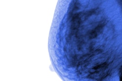
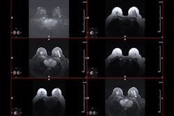
Contrast-enhanced mammography (CEM) can locally stage lobular breast carcinomas and could serve as an alternative to breast MRI, according to research published February 12 in Clinical Radiology.
A team led by Elisabetta Giannotti, MD, from Hospital NHS Foundation Trust in Cambridge, U.K., found that CEM had superior sensitivity compared to standard mammography and that both CEM and MRI overestimate tumor size to a similar degree.
"Traditionally, breast MRI has been used in many centers for preoperative staging, but CEM appears to be a suitable alternative,” the Giannotti team wrote.
Invasive lobular carcinoma is the second most common histological type of breast cancer after invasive ductal carcinoma. While it accounts for 5% to 15% of cases, previous reports suggest that the incidence of lobular carcinoma is increasing at a faster rate than other breast cancer subtypes.
The researchers also noted that ultrasound and mammography tend to underestimate lesion size in lobular carcinoma cases and that this type of breast cancer is less likely to be detected by mammography. The latter is due to the growth pattern of infiltration and density less than or equal to normal fibroglandular tissue. This makes breast MRI a recommended modality in preoperative workup in lobular carcinoma cases.
Previous studies suggest that CEM has comparable performance to breast MRI and could be helpful for patients with contraindications for MRI. Giannotti and colleagues studied CEM’s performance in the preoperative diagnosis and staging of invasive lobular carcinoma. They also compared the results to standard mammography and MRI.
The study included 115 lobular carcinoma cases, of which 46 (40%) presented symptomatically and 69 were detected by screening.
The correlation between the histological size measured on the surgical excision specimen size was greater with CEM than with standard mammography (r = 0.626 and 0.295 respectively, p = 0.001). Also, 19% of lobular carcinomas were not visible without the use of a contrast agent.
CEM also showed greater sensitivity (70%) for multifocal disease than that of standard mammography (20%, p < 0.0001). However, CEM was not superior in terms of specificity (82% and 98%, respectively, p = 0.035).
Additionally, CEM overestimated tumor size by an average of 1.5 times, which was comparable to MRI overestimating by an average of 1.3 times. On average, tumor sizes on CEM were 0.5 cm larger than those seen on MRI.
The study authors highlighted that their results aligned with those from previous single-center studies examining CEM for lobular carcinoma cases, which they called “very encouraging.”
“This concordance is important to confirm and corroborate the present independent conclusions,” the authors wrote.
The study can be found in its entirety here.