MRI-guided biopsies of non-mass enhancement breast lesions show significant malignancy rates, with women who have a known ipsilateral breast cancer showing much higher frequencies of invasive carcinoma (20% vs. 7%) and in situ carcinoma (27% vs. 9%) compared to women without prior breast cancer.
- Women with known ipsilateral malignancy had 20% invasive carcinoma rate compared to 7% in those without prior cancer
- In situ carcinoma frequency was 27% with ipsilateral malignancy versus 9% without
- Benign findings were significantly lower (46%) in patients with prior cancer versus 77% in those without
- Most invasive carcinomas were well to moderately differentiated and hormone receptor-positive
MRI-guided breast biopsies for non-mass enhancement (NME) lesions show a significant rate of malignancy, according to research published August 5 in Human Pathology.
Biopsies from women with a known ipsilateral malignancy have a higher frequency of invasive and in situ breast cancer, wrote a team led by Haiying Zhan, MD, PhD, from Yale University in New Haven, CT.
“NME on MRI in patients with a known ipsilateral breast cancer may warrant a lower threshold for biopsy, as additional foci of disease are more likely in this setting,” the Zhan team wrote.
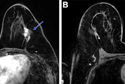
Breast MRI is recommended for screening women at high-risk of developing breast cancer. It is also used to examine disease extent in women with breast cancer. NME lesions can be challenging to interpret due to their broad radiologic–pathologic spectrum and variable association with malignancy, the researchers wrote.
Zhan and colleagues studied pathologic findings tied to MRI-guided core needle biopsies performed for NME breast lesions. The study included 270 MRI-guided core needle biopsies from 225 women performed in 2024 and 2025. All included cases showed NME on MRI.
The team divided these cases into two groups: women with a recent diagnosis of ipsilateral malignancy (n = 74) and women without a recent diagnosis of malignancy (n =196).
Biopsies from women with a known ipsilateral malignancy showed a higher frequency of invasive carcinoma and in situ carcinoma compared with women without ipsilateral malignancy. Women with a known ipsilateral malignancy also had a lower frequency of benign findings, the researchers observed.
Frequency of breast cancer in women with, without ipsilateral malignancy | |||
Measure | No ipsilateral malignancy | With ipsilateral malignancy | P value |
Frequency of invasive carcinoma | 7% | 20% | 0.003 |
Frequency of in situ carcinoma | 9% | 27% | 0.0002 |
Frequency of benign findings | 77% | 46% | 0.0001 |
Invasive carcinomas tied to NME included mostly well- to moderately-differentiated carcinomas (96%) and were ER–positive/HER2-negative (>90%).
Finally, ductal carcinoma in situ (DCIS) associated with NME included 57% high-grade and 43% intermediate-grade cases, with 36% showing an ER-negative staining.
The study authors highlighted their results in emphasizing the importance of clinical context in stratifying risk, since non-mass enhancements “may represent multifocal or multicentric disease.”
“Although hormone receptor-positive malignancies constitute the majority of cancers identified in NME, high-grade DCIS represents a meaningful subset and should remain a key consideration during clinical decision-making,” they wrote.
Read the full study here.