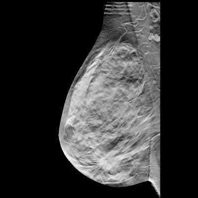
Single-view digital breast tomosynthesis (DBT) could use a little help from artificial intelligence (AI), according to research published July 6 in Radiology.
Researchers led by Maria Pinto from Radboud University in the Netherlands found that a single-view DBT and AI setup could allow for more effective screening with higher performance -- especially in terms of an increase in cancers detected -- than using single-view DBT alone.
The study authors said that radiologists improved their cancer detection performance and that the combined use of AI and DBT could allow for a more cost-effective screening program with higher performance and a lower workload for the radiologists.
Interpreting DBT images is challenging because of the higher volume of data that needs evaluation compared with interpreting conventional 2D digital mammography images. Pinto and colleagues wanted to see if adding AI to DBT could improve reading time and the performance of radiologists for breast cancer detection.
The team looked at breast exams acquired between June 2016 and February 2018 with a wide-angle DBT system. The median age of the patients was 54 years. Fourteen breast screening radiologists interpreted 190 DBT images from 190 women. Reading was performed in two sessions with a random mix of examinations being read with and without AI decision and navigation support.
The examination-based reader-averaged area under the curve was higher when interpreting results with AI support (0.88) than when reading unaided (0.85, p = 0.01). The average sensitivity increased with AI support (86%) than without it (81%, p = 0.006). The researchers found no statistically significant differences in specificity or reading time.
The team recommended that prospective, international, and multicenter screening studies be conducted to determine the impact of using AI-aided, single-view DBT for screening.

![A normal mammogram confirmed by three-year radiologic follow-up illustrates reader-marked regions of interest (ROIs) during (A) unaided (round 1) and (B) artificial intelligence (AI)–assisted (round 2) reading. Each colored dot represents an ROI for recall by a human reader. Readers could mark more than one ROI per case, represented by multiple dots of the same color. During AI-assisted reading, the AI system displayed three visible prompts: two with suspicion of malignancy scores of 35% (left mediolateral oblique [L MLO] and craniocaudal [L CC]) and one with a suspicion of malignancy score of 10% (right craniocaudal [R CC]), shown as polygonal overlays. Without AI, six of 10 readers (60%) marked a false-positive ROI. With AI assistance, this fell to two of 10 (20%). R MLO = right mediolateral oblique.](https://img.auntminnie.com/mindful/smg/workspaces/default/uploads/2026/07/2026-07-14-radiology-mammogram-ai-auto-bias.H0bYO8QlWs.jpg?auto=format%2Ccompress&fit=crop&h=100&q=70&w=100)


![A normal mammogram confirmed by three-year radiologic follow-up illustrates reader-marked regions of interest (ROIs) during (A) unaided (round 1) and (B) artificial intelligence (AI)–assisted (round 2) reading. Each colored dot represents an ROI for recall by a human reader. Readers could mark more than one ROI per case, represented by multiple dots of the same color. During AI-assisted reading, the AI system displayed three visible prompts: two with suspicion of malignancy scores of 35% (left mediolateral oblique [L MLO] and craniocaudal [L CC]) and one with a suspicion of malignancy score of 10% (right craniocaudal [R CC]), shown as polygonal overlays. Without AI, six of 10 readers (60%) marked a false-positive ROI. With AI assistance, this fell to two of 10 (20%). R MLO = right mediolateral oblique.](https://img.auntminnie.com/mindful/smg/workspaces/default/uploads/2026/07/2026-07-14-radiology-mammogram-ai-auto-bias.H0bYO8QlWs.jpg?auto=format%2Ccompress&fit=crop&h=112&q=70&w=112)