An AI approach could help improve workflows in screening digital breast tomosynthesis (DBT), according to findings published June 2 in Radiology.
The use of AI-based slab reconstruction technology -- where multiple thin image slices are combined into thicker composite images -- led to improved specificity and reduced abnormal interpretation rate while maintaining image quality, wrote a team led by Manisha Bahl, MD, from Massachusetts General Hospital in Boston.
“These findings suggest that slab reconstruction may help radiologists more confidently dismiss benign findings, potentially reducing false-positive assessments while preserving cancer detection,” Bahl told AuntMinnie.
With DBT resulting in more images to interpret than conventional mammography, radiologists are faced with heavier workloads and longer interpretation times. The standard image slice thickness on DBT is 1 mm, meaning a standard bilateral screening exam of a 50-mm breast may generate about 200 DBT images for review.
Slab reconstruction technologies aim to help with this by generating thicker image slices. This reduces the number of images per exam while preserving diagnostically relevant information.
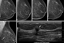
 Slab reconstruction provides visualization of subtle architectural distortion similar to that of standard 1-mm slices in a false-positive screening exam. Images include breast exams from a 45-year-old woman who underwent screening mammography. Craniocaudal tomosynthesis images with (A) 1-mm slices and (B) 6-mm slab reconstructions show an area of architectural distortion in the subareolar region of the left breast (arrow). (C) Targeted ultrasound imaging shows a corresponding irregular hypoechoic mass (arrow), which was subsequently biopsied. Pathologic examination revealed a sclerosing papillary lesion.Bahl et al/Radiology
Bahl and colleagues compared the performance of screening DBT before and after the implementation of AI-based slab reconstruction technology (3DQuorum, Hologic) that generates 6-mm synthetic slices with 3-mm overlap. The AI system also has a feature-preserving algorithm. The study included a pre-implementation period (2018 and 2019) and a post-implementation period (2021 and 2022).
Final analysis included 119,662 screening DBT exams in 64,949 women. Of the exams, 77,577 (in 52,649 women) took place during the pre-implementation period while 42,085 (in 42,059 women) occurred during the post-implementation period.
The AI approach improved specificity and abnormal interpretation rate while also resulting in noninferior cancer detection rate, false-negative rate, and sensitivity.
Comparison between pre-, post-AI implementation in screening DBT | ||||
Measure | Pre-AI | Post-AI | Odds ratio | P value |
Cancer detection rate (per 1,000 exams) | 5.8 | 6.5 | 1.1 | 0.5 |
Sensitivity | 82.3% | 85.9% | 1.3 | 0.3 |
Specificity | 94.4% | 94.9% | 1.1 | <0.001 |
False-negative rate (per 1,000 exams) | 1.2 | 1.1 | 0.8 | 0.4 |
Abnormal interpretation rate | 6.2% | 5.8% | 0.9 | <0.001 |
The findings support the use of slab-based DBT as a “safe and efficient alternative” in routine breast cancer screening, the study authors wrote. Bahl said this approach has the potential to improve screening workflow by decreasing image volume, reducing cognitive load, and supporting more efficient interpretation.
“Given the reduced image volume and maintained diagnostic performance observed in our study, these workflow benefits may be achievable without sacrificing diagnostic accuracy,” she added.
Bahl told AuntMinnie the research team is interested in better understanding the mechanisms underlying the improvements in specificity and abnormal interpretation rate observed in the study.
“Another important area for future research is how slab reconstruction interacts with newer AI tools for lesion detection and diagnostic decision support,” she said.
The findings provide “a promising new avenue” to improve efficiency for radiologists reading screening DBT exams, according to an accompanying editorial written by Lars Grimm, MD, from Duke University in Durham, NC.
“It remains to be seen if these results can translate to other DBT vendors and AI slab algorithms, as well as if the efficiency and performance gains persist with other AI tools such as CAD,” Grimm wrote. “Nevertheless, the breast imaging field as a whole is struggling to accommodate increasing volumes and workforce shortages, so efforts to meaningfully improve workplace efficiency can have far reaching beneficial effects.”
Read the full study here.
![A normal mammogram confirmed by three-year radiologic follow-up illustrates reader-marked regions of interest (ROIs) during (A) unaided (round 1) and (B) artificial intelligence (AI)–assisted (round 2) reading. Each colored dot represents an ROI for recall by a human reader. Readers could mark more than one ROI per case, represented by multiple dots of the same color. During AI-assisted reading, the AI system displayed three visible prompts: two with suspicion of malignancy scores of 35% (left mediolateral oblique [L MLO] and craniocaudal [L CC]) and one with a suspicion of malignancy score of 10% (right craniocaudal [R CC]), shown as polygonal overlays. Without AI, six of 10 readers (60%) marked a false-positive ROI. With AI assistance, this fell to two of 10 (20%). R MLO = right mediolateral oblique.](https://img.auntminnie.com/mindful/smg/workspaces/default/uploads/2026/07/2026-07-14-radiology-mammogram-ai-auto-bias.H0bYO8QlWs.jpg?auto=format%2Ccompress&fit=crop&h=100&q=70&w=100)


![A normal mammogram confirmed by three-year radiologic follow-up illustrates reader-marked regions of interest (ROIs) during (A) unaided (round 1) and (B) artificial intelligence (AI)–assisted (round 2) reading. Each colored dot represents an ROI for recall by a human reader. Readers could mark more than one ROI per case, represented by multiple dots of the same color. During AI-assisted reading, the AI system displayed three visible prompts: two with suspicion of malignancy scores of 35% (left mediolateral oblique [L MLO] and craniocaudal [L CC]) and one with a suspicion of malignancy score of 10% (right craniocaudal [R CC]), shown as polygonal overlays. Without AI, six of 10 readers (60%) marked a false-positive ROI. With AI assistance, this fell to two of 10 (20%). R MLO = right mediolateral oblique.](https://img.auntminnie.com/mindful/smg/workspaces/default/uploads/2026/07/2026-07-14-radiology-mammogram-ai-auto-bias.H0bYO8QlWs.jpg?auto=format%2Ccompress&fit=crop&h=167&q=70&w=250)
![A normal mammogram confirmed by three-year radiologic follow-up illustrates reader-marked regions of interest (ROIs) during (A) unaided (round 1) and (B) artificial intelligence (AI)–assisted (round 2) reading. Each colored dot represents an ROI for recall by a human reader. Readers could mark more than one ROI per case, represented by multiple dots of the same color. During AI-assisted reading, the AI system displayed three visible prompts: two with suspicion of malignancy scores of 35% (left mediolateral oblique [L MLO] and craniocaudal [L CC]) and one with a suspicion of malignancy score of 10% (right craniocaudal [R CC]), shown as polygonal overlays. Without AI, six of 10 readers (60%) marked a false-positive ROI. With AI assistance, this fell to two of 10 (20%). R MLO = right mediolateral oblique.](https://img.auntminnie.com/mindful/smg/workspaces/default/uploads/2026/07/2026-07-14-radiology-mammogram-ai-auto-bias.H0bYO8QlWs.jpg?auto=format%2Ccompress&dpr=2&fit=crop&h=167&q=70&w=250)