Attenuation imaging is feasible for ultrasonic detection of hepatic steatosis in patients with nonalcoholic fatty liver disease (NAFLD), according to a study published July 6 in Academic Radiology.
Researchers led by Yun-Lin Huang, MD, from Fudan University in Shanghai, China, found perfect examination success rate and high diagnostic performance marks for attenuation imaging, as well as comparable performance to controlled attenuation parameter ultrasound exams.
"Attenuation imaging measurement using an ultrasound scan system is relatively economical and timesaving, as it can detect the fat content of liver, as well as the hepatic morphology, echogenicity, and potential focal liver lesions," Huang and colleagues wrote.
Ultrasound is the recommended first-line screening tool for patients at risk of nonalcoholic fatty liver disease, with controlled attenuation parameter exams being used to quantify the amount of fat in the liver. This method measures fat content in the liver separately from fibrosis, making it a day-to-day clinical tool to measure hepatic steatosis.
However, this exam can only distinguish a little more than 10% of steatosis, according to the researchers. What's more, they noted that the technique cannot simultaneously evaluate morphological changes in the liver, and they also perform poorly in patients with higher body mass index (BMI) levels.
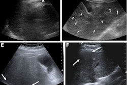
The researchers hypothesized that attenuation imaging could help overcome these shortcomings. This method quantitatively detects the degree of ultrasonic acoustic wave attenuation. It also has a larger sampling box and conducts a morphological evaluation by dual imaging with B-mode ultrasound and attenuation imaging.
Huang and colleagues wanted to investigate this method's performance for detecting and quantifying hepatic steatosis in patients who had clinical suspicion of non-alcoholic fatty liver disease. They used histopathology as the reference standard.
The study included 60 patients who were referred for liver biopsy after attenuation imaging and controlled attenuation parameter examinations in 2020 and 2021.
The researchers reported a 100% success rate for the attenuation imaging exam, as well as an intraobserver reproducibility of 0.981. They also found comparable area under the curve (AUC) values between attenuation imaging and controlled attenuation parameter exams.
| AUC of ultrasound imaging methods for detecting hepatic steatosis in NAFLD | |||
| Fatty liver stage | Controlled attenuation parameter | Attenuation imaging | p-value |
| Stage 1 | 0.916 | 0.968 | 0.281 |
| Stage 2 | 0.872 | 0.911 | 0.254 |
| Stage 3 | 0.807 | 0.766 | 0.33 |
The team also reported that attenuation imaging had significant correlations with high-density lipoprotein cholesterol (p < 0.001), and with triglycerides (p = 0.015).
Huang et al highlighted that attenuation imaging's advantage is that it evaluates liver fat with a color-coded map overlaid on B-mode ultrasound images. They also wrote that the average time of the attenuation imaging exams was 3.8 minutes, suggesting that radiologists "can easily measure attenuation imaging values through the automatic removal of vessels and artifacts and the display of the reliability index."
The authors called for their results to be confirmed by future research with large sample sizes in a multicenter study.
The study can be found in its entirety here.