Certain ultrasound parameters have high diagnostic value in detecting liver steatosis and fibrosis, making way for attenuation imaging and shear-wave elastography, a study published July 26 in Academic Radiology found.
Researchers led by Mehnoosh Torkzaban, MD, from Thomas Jefferson University in Philadelphia found that attenuation coefficient and liver stiffness showed high marks in detecting these liver diseases. They also found that combining these parameters further improved detection of nonalcoholic fatty liver disease and patients at higher risk for this disease.
"This study showed that attenuation imaging and shear wave elastography are useful ultrasound tests for detecting steatosis and fibrosis among nonalcoholic fatty liver disease suspects, respectively," Torkzaban and colleagues wrote.
Marked liver steatosis, steatohepatitis, and significant fibrosis are established risk factors for poor outcomes in nonalcoholic fatty liver disease. New innovations in ultrasound imaging have been investigated in this setting, with previous research suggesting strong correlations between imaging parameters and liver activity.
Torkzaban and colleagues wanted to investigate quantitative ultrasound parameters and compare their diagnostic value to that of standard of care, including liver biopsy and MRI. The researchers prospectively studied the performance of attenuation coefficient, liver stiffness, and dispersion slope in diagnosing liver steatosis, fibrosis, and inflammation, respectively. They also assessed the feasibility of predicting clinically significant fibrosis (stage ≥ 2) by using combined ultrasound parameters in nonalcoholic fatty liver disease patients.
The team included a total of 74 patients, 34 of whom underwent liver biopsy and the other 40 undergoing MRI. Also, 32 patients had liver steatosis and fibrosis, 22 had steatosis, five had fibrosis, and 15 had normal livers.
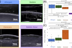
The researchers found significantly higher average attenuation coefficients in steatotic livers (n = 54) than in nonsteatotic livers (n = 20, p < 0.0001). They also found significantly higher liver stiffness and dispersion slope values in patients with liver fibrosis compared with nonfibrotic livers (p = 0.0004 and p = 0.0002, respectively).
The team also found area under the receiver operating characteristic curve (AUROCC) values of 0.87 for combined ultrasound parameters of liver stiffness and attenuation coefficient in detecting liver steatosis and fibrosis. This included a negative predictive value (NPV) of 75% and a positive predictive value (PPV) of 77% (p < 0.0001). Additionally, liver stiffness showed an AUROCC of 0.93 in detecting patients with liver steatosis and fibrosis stage ≥ 2, including a negative predictive value (NPV) of 87% and a positive predictive value (PPV) of 82% (p < 0.0001).
The researchers also found that for the patients who underwent biopsy, 11 were diagnosed with nonalcoholic steatohepatitis. Dispersion slope values indicated a significant difference between patients with (n = 23) or without (n = 11) hepatocellular ballooning (p = 0.02).
Finally, the team reported that when all three ultrasound parameters were combined with body mass index, the AUROCC for detecting nonalcoholic steatohepatitis was 0.87. This included a NPV of 80% and a PPV of 87% (p = 0.0006).
The study authors suggested that based on their results, attenuation imaging and shear-wave elastography could serve as potential screening or monitoring tools for these patients.
However, they also called for future studies to include large, diverse populations to find out the best combination of ultrasound parameters for detecting nonalcoholic steatohepatitis. This includes determining cutoff values comparable to different stages of fibrosis, steatosis, and inflammation.
The study can be found in its entirety here.