Models that use quantitative ultrasound parameters showed reliable performance for discriminating steatosis in patients with chronic liver disease, according to results published October 3 in Radiology.
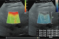
Researchers led by Hidekatsu Kuroda, MD, PhD, from Iwate Medical University School of Medicine in Japan found that one of their models using integrated backscatter coefficient (IBSC), signal-to-noise ratio (SNR), and ultrasound-guided attenuation parameter (UGAP) can accurately discriminate steatosis of at least 5% in chronic liver disease patients.
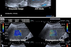
“Our results suggest that composite evaluations of ultrasound radiofrequency signals can identify biologic tissue characteristics of liver steatosis,” Kuroda and colleagues wrote.
Previous reports suggest an increase worldwide in the incidence of nonalcoholic fatty liver disease. The researchers noted that this means noninvasive, widely available methods are needed for accurately assessing liver steatosis.
The Kuroda team sought to investigate the performance of models using different combinations of quantitative ultrasound parameters. Specifically, it wanted to find the models’ ability to predict at least 5% steatosis in patients as defined using MRI proton density fat fraction. The group used four models for the study: model 1 (UGAP alone), model 2 (UGAP with IBSC), model 3 (UGAP with SNR), and model 4 (UGAP with IBSC and SNR).
The researchers included 582 participants with a median age of 64 years in their study. Of these, 364 were placed into the steatosis group and 218 were in the nonsteatosis group.
The researchers found that while all models demonstrated high area under the curve (AUC) values, model 4 with all parameters included had the highest AUC value and the highest C-index of all models adjusted for bootstrap analysis, which resamples data to create many simulated samples.
| Comparison of models using ultrasound parameters | ||||
|---|---|---|---|---|
| Model 1 | Model 2 | Model 3 | Model 4 | |
| AUC | 0.92 | 0.93 | 0.95 | 0.96 |
| C-index | 0.92 | 0.93 | 0.95 | 0.96 |
The team also reported that compared with other models, models 3 and 4 showed statistically significant improvement in discriminating at least 5% steatosis (p < .01).
While the study authors suggested that these parameters as well as others may be more appropriate for ultrasonic diagnosis of steatosis, more research is needed to “create a more promising” diagnostic approach.
“In the future, external validation studies are needed to determine whether these findings can be widely applied in primary care settings,” they wrote.
In an accompanying editorial, Aiguo Han, PhD, from the Virginia Polytechnic Institute and State University wrote that some future directions in this area include translating the use of this technology from adults to children, developing vendor-independent methods, and investigating factors that may impact parameter measurements, among others.
“This study is an important step toward a more accurate quantitative ultrasound-based assessment of hepatic steatosis,” Han wrote.
The full study can be found here.