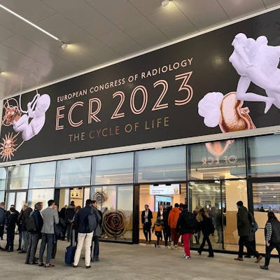
Superb microvascular imaging (SMI) with elastosonography may not be as valuable as standard TI-RADS, but it does hold some potential, according to findings presented March 1 at ECR 2023.
In his talk, Dr. Davide Negroni from Maggiore Hospital in Novara, Italy discussed his team's findings, showing that this combined method found solid nodules appearing to be at greater risk of malignancy. However, it overall had a lower diagnostic performance than TI-RADS as defined by the American College of Radiology (ACR TI-RADS).
"TI-RADS is the most predictive score for malignancy, compared with SMI and elastography," Negroni said.
Previous data suggests about a 50% prevalence of thyroid nodules larger than one cm in patients without previously diagnosed thyroid disease. While most detected nodules are benign and clinically insignificant, Negroni said that thyroid nodules are clinically important as they may indicate thyroid cancer in 4% to 6.5% of cases.
Ultrasound is the gold standard modality in detecting these nodules. However, alternative methods to conventional ultrasound have been explored in recent years by researchers.
 Dr. Davide Negroni presents findings from research he led on combining SMI with elastosonography for diagnosing thyroid lesions. While the method's diagnostic performance was lower than ACR TI-RADS when used in a heterogeneous, it may yield some potential.
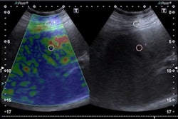
SMI helps visualize low velocity and microvascular flow by using a clutter suppression algorithm. From there, it provides color overlay images, with previous studies suggesting it can provide additional value over conventional color or power Doppler imaging. Elastography meanwhile measures stiffness of tissues, with harder tissues indicating potential malignancies.
Negroni et al wanted to look at the performance of SMI and elastography in comparison with ACR TI-RADS when it comes to diagnosing thyroid nodules in a heterogeneous population.
They looked at a total of 260 thyroid nodules from 251 consecutive patients with an average age of 58.6 years. The patients presented with at least one thyroid nodule and were candidates for fine-needle aspiration. All identified nodules underwent fine-needle aspiration, and the examination reported a success rate of 86.5% of the sampling. Most of the nodules found were either TI-RADS 3 or 4 (69.2%).
The researchers found that area under the curve (AUC) values favored ACR TI-RADS over SMI, elastography, or combined SMI and elastography.
| Diagnostic performance on thyroid nodules (AUC) | |
| ACR TI-RADS | 0.673 |
| SMI plus elastography | 0.616 |
| SMI | 0.576 |
| Elastography | 0.576 |
By combining all three standalone methods, the AUC improved to 0.699. Additionally, owing to elastography, solid nodules on thyroid findings appear at greater risk of malignancy, Negroni said. He added that no specific vascular patterns at SMI of evolving lesions were identified.
Negroni also reported that SMI adds about one to two minutes to the total image acquisition time, though using the "strain" technique for elastography adds five to six minutes.
"Usually, I have about 32 thyroid searches every day, it would be quite difficult to use this in every patient," he said.