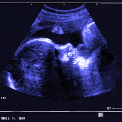
Cervical changes can be seen between pregnant and nonpregnant women via strain elastography, according to findings presented March 28 at UltraCon.
Presenter Dr. Nora Doyle from University of Texas Health Science Center at San Antonio shared her team's findings, which showed significant changes in cervical tissue consistency and density when comparing the pregnant and nonpregnant cervix.
"Practitioners may be able to use this as a screening tool for preterm birth and other interventions like cerclage placement to save a pregnancy," Doyle told AuntMinnie.com.
Transvaginal ultrasound (TVUS) is commonly used to look at cervical changes throughout a pregnancy. Previous studies have suggested that elastography is useful in assessing human tissues in several medical specialties, but Doyle noted that it hasn't been used much in obstetrics.
The researchers sought to compare the tissue consistency differences in pregnant and nonpregnant cervixes using strain elastography. They assessed 111 cervical measurements collected between 2019 and 2020; of these, 68 were gathered from pregnant women and 43 from nonpregnant women. For the pregnant women, 55 were singleton and 13 were twin pregnancies.
The team found significant differences between pregnant and nonpregnant cervixes in several measurements.
| Comparison between pregnant, nonpregnant women | ||
| Measurement | Nonpregnant women | Pregnant women |
| Cervical length | 2.83 cm | 3.83 cm |
| Hardness ratio | 91.3% | 59.5% |
| Elasticity contrast index | 2.73 | 4.17 |
| Internal Os strain | 0.12 | 0.28 |
| External Os strain | 0.13 | 0.31 |
The researchers also noted that these differences held true through all pregnancy trimesters. They also found no significant difference in the ratio of internal os strain and external os strain between pregnant and nonpregnant women (p = 0.99).
The study showed that women pregnant with twins had cervical tissue that was less dense than cervical tissue for those with a singleton pregnancy and for women who were not pregnant. This included an average cervical length of 4.09 for twin pregnancies, as well as a hardness ratio of 57.1%.
However, the team found no significant difference in the ratio of internal os strain and external os strain between women carrying a singleton pregnancy and nonpregnant women, or between women carrying twins and nonpregnant women.
Doyle's group plans to more data to construct a cervical nomogram in pregnancy, she said.


![Examples of ultrasound findings and techniques. (A) Images in a 39-year-old male patient with a mass in the left thigh. The mass is heterogeneous on the B-mode US image (compared with the patient in D) and showed increased microvascularity (superb microvascular imaging [SMI]) and shear-wave elastography (SWE) values. Undifferentiated pleomorphic sarcoma was diagnosed at biopsy (with pleomorphic rhabdomyosarcoma in surgical specimen). (B) Images in an 18-year-old male patient with a mass in the left leg. The mass is hypoechoic on the B-mode image, with no other findings suggestive of malignancy. The lesion is in contact with the cortex of the tibia, which is slightly irregular. CT revealed a doubtful anteromedial tibial erosion. The microvascular study demonstrated high vascularization, suggestive of malignancy. Periosteal Ewing sarcoma was diagnosed with both histologic and immunohistochemical confirmation. (C) Images in a 69-year-old female patient with a lump growing on the outside of the left leg. Multiple SWE examinations were performed (please note the high values obtained in the measurements, whereas the color map highlights the stiffness relative to adjacent tissues). SMI showed areas of increased vascularization to target for sampling. Undifferentiated spindle cell sarcoma was diagnosed at biopsy, with residual leiomyosarcoma in the surgical specimen after neoadjuvant therapy. (D) Images in a 56-year-old female patient with a mass in the right thigh. The mass is heterogeneous at both B-mode ultrasound (similar to patient A) and MRI (coronal T2-weighted spectral attenuated inversion recovery [SPAIR]; T1-weighted pre-contrast and postcontrast imaging), which even shows uptake after the administration of paramagnetic contrast material, which is traditionally suggestive of malignancy. Low values at SMI and elastography are suggestive of benignity. Spindle cell lipoma was diagnosed at biopsy, with atypical spindle cell lipomatous tumor in the surgical specimen.](https://img.auntminnie.com/mindful/smg/workspaces/default/uploads/2026/08/images-radiol250278fig2.APCFLSvX6p.jpg?auto=format%2Ccompress&fit=crop&h=100&q=70&w=100)


![Examples of ultrasound findings and techniques. (A) Images in a 39-year-old male patient with a mass in the left thigh. The mass is heterogeneous on the B-mode US image (compared with the patient in D) and showed increased microvascularity (superb microvascular imaging [SMI]) and shear-wave elastography (SWE) values. Undifferentiated pleomorphic sarcoma was diagnosed at biopsy (with pleomorphic rhabdomyosarcoma in surgical specimen). (B) Images in an 18-year-old male patient with a mass in the left leg. The mass is hypoechoic on the B-mode image, with no other findings suggestive of malignancy. The lesion is in contact with the cortex of the tibia, which is slightly irregular. CT revealed a doubtful anteromedial tibial erosion. The microvascular study demonstrated high vascularization, suggestive of malignancy. Periosteal Ewing sarcoma was diagnosed with both histologic and immunohistochemical confirmation. (C) Images in a 69-year-old female patient with a lump growing on the outside of the left leg. Multiple SWE examinations were performed (please note the high values obtained in the measurements, whereas the color map highlights the stiffness relative to adjacent tissues). SMI showed areas of increased vascularization to target for sampling. Undifferentiated spindle cell sarcoma was diagnosed at biopsy, with residual leiomyosarcoma in the surgical specimen after neoadjuvant therapy. (D) Images in a 56-year-old female patient with a mass in the right thigh. The mass is heterogeneous at both B-mode ultrasound (similar to patient A) and MRI (coronal T2-weighted spectral attenuated inversion recovery [SPAIR]; T1-weighted pre-contrast and postcontrast imaging), which even shows uptake after the administration of paramagnetic contrast material, which is traditionally suggestive of malignancy. Low values at SMI and elastography are suggestive of benignity. Spindle cell lipoma was diagnosed at biopsy, with atypical spindle cell lipomatous tumor in the surgical specimen.](https://img.auntminnie.com/mindful/smg/workspaces/default/uploads/2026/08/images-radiol250278fig2.APCFLSvX6p.jpg?auto=format%2Ccompress&fit=crop&h=112&q=70&w=112)