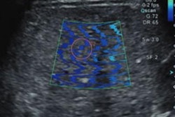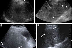
Ultrasonic strain elastography shows higher liver stiffness measures in recovered long COVID-19 patients, according to research presented at ECR 2023.
Dr. Yusuf Aksu from Necip Fazıl City Hospital in Kahramanmaras, Turkey, shared findings from a study he and colleagues conducted that showed significantly higher liver strain ratios in COVID-19 patients than in healthy volunteers who never had COVID-19.
"Our results showed a continuing increasing of liver stiffness in post-long COVID-19 patients," he told session attendees.
Patients with COVID-19 tend to present with symptoms of viral pneumonia or multisystem inflammatory disorders, and the liver is one of the organs most affected, Aksu said. As COVID-19 runs its course, liver enzymes may be elevated and liver insufficiency can develop.
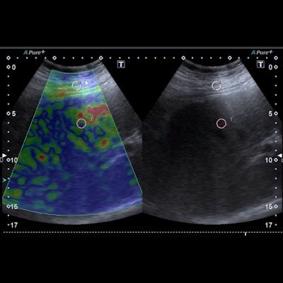
Ultrasonic elastography can measure tissue stiffness caused by a variety of diseases, Aksu noted, and strain elastography is a qualitative method that provides information on the relative stiffness between one tissue and another. The technique differs from shear-wave elastography, which uses shear-wave speed to estimate tissue stiffness values.
 Dr. Yusuf Aksu from Necip Fazıl City Hospital in Kahramanmaras, Turkey presents findings at ECR 2023 that demonstrate how strain elastography shows higher liver stiffness in patients who have recovered from long COVID-19.
Previous studies have suggested that elevated liver strain ratio during acute COVID-19 reflects systemic or liver involvement, according to Aksu, who noted that elevated liver strain ratio can indicate clinical worsening of COVID-19 patients.
Aksu's group sought to evaluate liver stiffness changes with strain elastography in long COVID-19 patients who had completely recovered and had no comorbidities. The team conducted a study that included data collected in 2022 from 175 patients who were split into two groups: a post-long COVID-19 group (91) and a healthy volunteer group (84). The researchers took strain elastography measurements from the left, right, and caudate liver lobes.
They found that average liver strain ratio was significantly higher in the COVID group, at 1.00 compared to 0.72 in the healthy group (p < 0.0001). As for the receiver operating characteristic (ROC) curve, the team discovered that the optimum cutoff value for liver strain ratio in the COVID group was 1.31 -- which translated to a sensitivity of 38.5%, a specificity of 86.9%, and an area under the curve (AUC) of 0.646 (p < 0.001). The investigators found no significant differences in either group when it came to demographic characteristics and body mass index (BMI).
More prospective studies should be performed to further measure COVID-19's long-term effects, Aksu concluded.