Panoramic ultrasound does not produce imaging deformations during assessment of large muscles and shows good comparison with conventional B-mode ultrasound, suggests a study published January 10 in Physica Medica.
Researchers led by Juan Antonio Valera-Calero, PhD, from Complutense University of Madrid, Spain, found good to excellent agreement between panoramic and B-mode ultrasound methods when it came to assessing cross-sectional area, perimeter, circularity, aspect ratio, roundness, and average echogenicity of neck and short rotator muscles.
"Panoramic ultrasound could be used for research purposes in both cross-sectional and longitudinal designs to obtain reliable size, shape, and brightness scores," Valera-Calero and colleagues wrote.
Panoramic ultrasound offers a wider field of view than standard B-mode ultrasound is able to obtain. For example, panoramic ultrasound can display both thyroid gland lobes on a single image, which could further help in diagnosing diseases and disorders. However, for this method to be successful, transducer movement and speed must be constant to avoid imaging deformations, according to the researchers.
Valera-Calero and co-authors wanted to compare several morphology indicators and average echointensity in muscles via panoramic and conventional B-mode ultrasound images. For the study, the team included 46 healthy volunteers and used ultrasound imaging on the cervical multifidus muscle and short rotator muscles. The indicators the team looked at included area, perimeter, circularity, aspect ratio, and roundness.
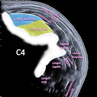
 B-mode ultrasound imaging shows short rotator (yellow) and cervical multifidus (blue) muscles. Panoramic ultrasound imaging of posterior neck muscles shows short rotators (yellow) and cervical multifidus (blue). Images courtesy of Physica Medica under CC BY 4.0 International license.
The researchers found that all parameters studied demonstrated no significant differences between both ultrasound methods for both muscles examined (p > 0.05 for all).
They also reported that panoramic ultrasound showed "excellent" agreement with B-mode ultrasound when analyzing all parameters for the cervical multifidus muscle, with intraclass correlation coefficient (ICC) values above 0.9. For short rotator muscles, the agreement ranged from good to excellent, with ICC values ranging from 0.861 to 0.978.
The study authors suggested that based on their results, panoramic ultrasound could serve as an accessible, low-cost imaging tool.
"In addition, our results could be interpreted as a methodological quality reinforcement of previous research using panoramic ultrasound imaging," they wrote.
They added that this could especially go for research exploring the same muscle regions the researchers analyzed in this most recent study, where "bias related with muscle deformations during the imaging acquisition could be suspected."
The authors noted that their findings should now be confirmed by comparing parameters obtained by panoramic ultrasound results with those obtained by MRI or CT.