Researchers recently developed a technique that creates an ultrasound "tornado" in order to treat acute blood clots inside the brain. Preliminary testing shows promising results in patients with a cerebral venous sinus thrombosis (CVST).
The investigators, led by Bohua Zhang, PhD, from North Carolina State University, found that their "vortex ultrasound" method could break down clots faster than conventional techniques in an in vitro, proof-of-concept model. Their results were published January 11 in Research.
"In a 3D cerebral venous sinus phantom, we found that we can recanalize an acute blood clot with eight minutes of treatment to restore the blood flow," wrote corresponding author Dr. Chengzhi Shi, PhD, from the Georgia Institute of Technology in Atlanta. "In addition, we found that the vortex ultrasound treatment is safe to the blood vessel."
CVST occurs when a blood clot forms in the veins responsible for draining blood from the brain. Although this incidence isn't common, between two and three per 100,000 cases per year in the U.S., the researchers noted that this trend is rising. They added that current treatment methods fail in between 20% and 40% of cases.
Zhang and co-authors wanted to show the effectiveness of a vortex ultrasound approach in treating acute blood clots in blood vessels leading to the brain, using a tool leveraging ultrasound transducer array technology.
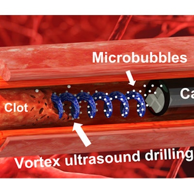
The tool is delivered via a catheter into the circulatory system and from there, into the blood clot. Inside the catheter is a single transducer designed to produce a swirling, vortex effect, such as that seen in a tornado. The idea is that this vortex ultrasound approach produces an in-plane pressure gradient, causing localized shear stress in the blood clot. For their in vitro, 3D-printed brain model, the researchers used cow blood.
 Researchers have developed a new tool and technique that uses "vortex ultrasound" -- a sort of ultrasonic tornado -- to break down blood clots in the brain. The new approach worked more quickly than existing techniques to eliminate clots formed in an in vitro model of cerebral venous sinus thrombosis (CVST). Image courtesy of North Carolina State University.
The team found that the lytic rate -- the degeneration of blood clots -- improved by at least 64.3% compared with nonvortex endovascular ultrasound treatment. The group also found that a 3.1 g, 7.5 cm long, completely blocked model of acute CVST saw restored blood flow within eight minutes. This included a record-high lytic rate of 237.5 mg per minute for acute bovine clot in vitro.
"Patients with CVST will usually have pressure build up in the vessel and the local hypertension can rupture the vessel wall and lead to fatal intracerebral hemorrhage," Shi told AuntMinnie.com. "So, time is life for the patients."
The researchers also highlighted that an acute blood clot in their model could be dissolved by the ultrasound tool in under half an hour. For comparison, conventional treatment methods take "at least" 15 hours -- over a day on average. The team also reported no damage to blood vessel walls inside the brain, as well as no "substantial" damage to red blood cells.
"This device was the first to incorporate the novel contrast agent-mediated vortex ultrasound for clot-dissolving technology into a 9-French catheter device to demonstrate the remarkably increased lytic efficiency and safety over existing thrombolysis approaches," the authors wrote.
The researchers indicated that the next step is conducting animal trials and then, if successful, launching clinical trials. They also suggested that if the tool ever becomes a clinical application, it would have comparable cost to conventional CVST interventions.
For future studies, the authors wrote that other parameters to consider in this approach include concentration of microbubbles, flow rate for microbubble injection, shear stress, and dosage for tissue plasminogen activators.