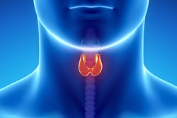
American Thyroid Association (ATA) guidelines are more effective than the American College of Radiology Thyroid Imaging Reporting and Data System (ACR-TIRADS) when evaluating pediatric thyroid nodules with ultrasound, a group of researchers has found.
The results could improve thyroid cancer management and care in children, who are vulnerable to being diagnosed at later stages and thus to poorer outcomes, wrote a team led by Dr. Gabriel Dunya from Lebanese American University-Rizk Hospital in Beirut. Dunya's and colleagues' findings were published December 12 in the International Journal of Pediatric Otorhinolaryngology.
"Early detection and appropriate management of pediatric thyroid nodules are of paramount importance, given that pediatric thyroid cancer is more likely to have an extrathyroidal extension and metastases at time of diagnosis," the group noted.
While new cases of thyroid cancer in younger people represent only 2.3% of all thyroid malignancies, reports suggest the rate is increasing, the investigators wrote. Early detection is key to managing cancer for better prognosis, and ultrasound is the standard modality for detecting thyroid nodules, with TIRADS and ATA guidelines being the most common stratification systems used.
Yet there are differences between the two guidelines, particularly regarding fine-needle aspiration. For example, ATA guidelines state that in children with non-hyperfunctioning thyroid nodules detected on ultrasound, fine-needle aspiration should be performed for all solid or part-solid nodules greater than 1 cm regardless of imaging features and that for children with suspicious thyroid nodules, a comprehensive cervical lymph node ultrasound should be performed, and all fine-needle aspirations should be guided by ultrasound. TI-RADS does not include this particular language.
Dunya's team sought to compare the two guidelines to determine which set is most sensitive based on final pathology outcomes. The study included 77 pediatric patients, all of whom underwent fine-needle aspiration as recommended by ATA guidelines. Of the lesions identified, 54.5% were benign, 22.1% were suspicious, and 23.4% were malignant.
Overall, Dunya's group found that the ATA guidelines yielded a sensitivity of 100% for identifying thyroid nodules, while the TIRADS system had a sensitivity rate of 75%.
When the study authors analyzed the data using the TIRADS guideline, they reported that 40.5% of these patients would have skipped a necessary fine-needle aspiration, while all malignant lesions would have undergone aspiration under ATA guidelines. The group also found that by using TIRADS, 5.6% of malignant lesions would have been missed.
More research is needed to compare the two frameworks, according to the authors.
"We encourage future prospective studies that compare both criteria in order to strengthen the results and increase the using of ATA guidelines," they concluded.



![Examples of ultrasound findings and techniques. (A) Images in a 39-year-old male patient with a mass in the left thigh. The mass is heterogeneous on the B-mode US image (compared with the patient in D) and showed increased microvascularity (superb microvascular imaging [SMI]) and shear-wave elastography (SWE) values. Undifferentiated pleomorphic sarcoma was diagnosed at biopsy (with pleomorphic rhabdomyosarcoma in surgical specimen). (B) Images in an 18-year-old male patient with a mass in the left leg. The mass is hypoechoic on the B-mode image, with no other findings suggestive of malignancy. The lesion is in contact with the cortex of the tibia, which is slightly irregular. CT revealed a doubtful anteromedial tibial erosion. The microvascular study demonstrated high vascularization, suggestive of malignancy. Periosteal Ewing sarcoma was diagnosed with both histologic and immunohistochemical confirmation. (C) Images in a 69-year-old female patient with a lump growing on the outside of the left leg. Multiple SWE examinations were performed (please note the high values obtained in the measurements, whereas the color map highlights the stiffness relative to adjacent tissues). SMI showed areas of increased vascularization to target for sampling. Undifferentiated spindle cell sarcoma was diagnosed at biopsy, with residual leiomyosarcoma in the surgical specimen after neoadjuvant therapy. (D) Images in a 56-year-old female patient with a mass in the right thigh. The mass is heterogeneous at both B-mode ultrasound (similar to patient A) and MRI (coronal T2-weighted spectral attenuated inversion recovery [SPAIR]; T1-weighted pre-contrast and postcontrast imaging), which even shows uptake after the administration of paramagnetic contrast material, which is traditionally suggestive of malignancy. Low values at SMI and elastography are suggestive of benignity. Spindle cell lipoma was diagnosed at biopsy, with atypical spindle cell lipomatous tumor in the surgical specimen.](https://img.auntminnie.com/mindful/smg/workspaces/default/uploads/2026/08/images-radiol250278fig2.APCFLSvX6p.jpg?auto=format%2Ccompress&fit=crop&h=100&q=70&w=100)


![Examples of ultrasound findings and techniques. (A) Images in a 39-year-old male patient with a mass in the left thigh. The mass is heterogeneous on the B-mode US image (compared with the patient in D) and showed increased microvascularity (superb microvascular imaging [SMI]) and shear-wave elastography (SWE) values. Undifferentiated pleomorphic sarcoma was diagnosed at biopsy (with pleomorphic rhabdomyosarcoma in surgical specimen). (B) Images in an 18-year-old male patient with a mass in the left leg. The mass is hypoechoic on the B-mode image, with no other findings suggestive of malignancy. The lesion is in contact with the cortex of the tibia, which is slightly irregular. CT revealed a doubtful anteromedial tibial erosion. The microvascular study demonstrated high vascularization, suggestive of malignancy. Periosteal Ewing sarcoma was diagnosed with both histologic and immunohistochemical confirmation. (C) Images in a 69-year-old female patient with a lump growing on the outside of the left leg. Multiple SWE examinations were performed (please note the high values obtained in the measurements, whereas the color map highlights the stiffness relative to adjacent tissues). SMI showed areas of increased vascularization to target for sampling. Undifferentiated spindle cell sarcoma was diagnosed at biopsy, with residual leiomyosarcoma in the surgical specimen after neoadjuvant therapy. (D) Images in a 56-year-old female patient with a mass in the right thigh. The mass is heterogeneous at both B-mode ultrasound (similar to patient A) and MRI (coronal T2-weighted spectral attenuated inversion recovery [SPAIR]; T1-weighted pre-contrast and postcontrast imaging), which even shows uptake after the administration of paramagnetic contrast material, which is traditionally suggestive of malignancy. Low values at SMI and elastography are suggestive of benignity. Spindle cell lipoma was diagnosed at biopsy, with atypical spindle cell lipomatous tumor in the surgical specimen.](https://img.auntminnie.com/mindful/smg/workspaces/default/uploads/2026/08/images-radiol250278fig2.APCFLSvX6p.jpg?auto=format%2Ccompress&fit=crop&h=112&q=70&w=112)