Schwannoma:
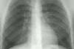
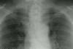
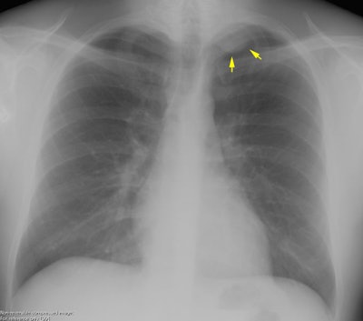
The patient shown below was asymptomatic and had his chest radiograph performed as part of a routine physical exam. The chest radiograph demonstrated a left apical soft tissue density mass (yellow arrows) with smooth inferior margins. A CT scan was performed (see below).
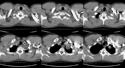
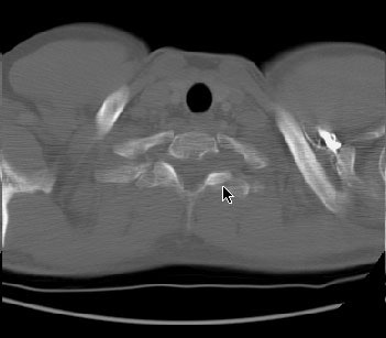
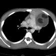
The CT scan revealed a homogeneous low density mass within the left lung apex- on careful evaluation, the mass could be seen extending outside the thoracic cavity and into the T1 neural foramena (middle image on top row and bone window shown below)
