Radiology 1996 Nov;201(2):471-474. Thymus in myasthenia gravis: comparison of CT and pathologic findings and clinical outcome after thymectomy.
Nicolaou S, Muller NL, Li DK, Oger JJ
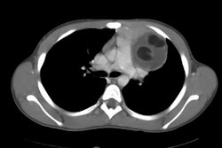
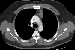
PURPOSE: To correlate computed tomographic (CT) appearance of the thymus with results from histologic examination of thymic tissue and clinical outcome in patients with generalized myasthenia gravis who underwent thymectomy. MATERIALS AND METHODS: Forty-five patients with myasthenia gravis underwent CT of the thorax and thymectomy. Findings at clinical follow-up were available in all patients. RESULTS: Twenty-six patients had normal CT findings, seven had a diffusely enlarged thymus, and 12 had a focal mass. The results of histologic examination showed that 16 of 26 patients with normal CT findings had normal thymic tissue and 10 had lymphoid follicular hyperplasia; all seven patients with an enlarged thymus had lymphoid hyperplasia. Five of 12 patients with a focal mass at CT had lymphoid hyperplasia, and seven had thymoma. Clinical improvement following thymectomy was observed in 27 (93%) of 29 patients with lymphoid hyperplasia or thymoma and 11 (69%) of 16 patients with normal histologic examination (P < .03, chi(2) test). CONCLUSION: The presence of an enlarged thymus or a focal mass in patients with myasthenia gravis indicates lymphoid hyperplasia or thymoma. However, CT is of limited value in distinguishing lymphoid follicular hyperplasia from a normal thymus or thymoma and in predicting clinical outcome.
PMID: 8888243, MUID: 97043045