Mediastinal teratoma: CT differentiation of ruptured and unruptured tumors.
Choi SJ, Lee JS, Song KS, Lim TH
OBJECTIVE: The purpose of this study was to differentiate ruptured from
unruptured mediastinal teratomas using CT. MATERIALS AND METHODS; CT findings
in 17 cases of surgically resected mediastinal teratomas were reviewed
retrospectively. Preoperative rupture was found in seven patients during
surgery. We compared the clinical symptoms and CT findings of ruptured
tumors with those of unruptured tumors. On CT, we evaluated size, wall
thickness, location of the mass, presence or absence of internal septation,
homogeneity of the internal components of each compartment, calcification
or fat within the mass, and ancillary findings in adjacent structures.
RESULTS: Severe symptoms (chest pain or hemoptysis) were more commonly
found in ruptured (71%) than in unruptured tumors. All ruptured mediastinal
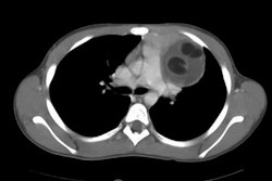
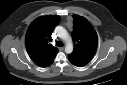
teratomas had a tendency to display inhomogeneity of the internal components,
whereas 90% of unruptured masses showed homogeneous densities of internal
components in each compartment of the mass. Ancillary CT findings in ruptured
tumors included fat-containing masses in adjacent lung parenchyma in two
patients, consolidation or atelectasis in the adjacent lung in three patients,
pericardial effusion in one patient, and pleural effusion in four patients.
CONCLUSION: In cases of mediastinal teratoma, CT findings of inhomogeneity
of the internal components and changes in the adjacent lung parenchyma,
pleura, or pericardium can be used as signs of tumor rupture.