Pericardial Cyst:
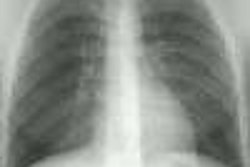
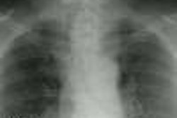
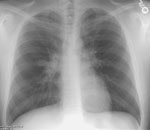
The patient shown below presented for routine chest radiograph prior to orthopedic surgery.
(Click on the small images to view the larger radiographs)
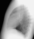
The PA exam revealed right paratracheal and azygous nodal soft tissue fullness (red
arrows). The lateral exam is also provided.
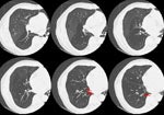
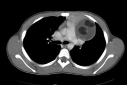
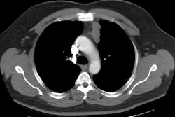
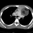
The CT scan demonstrated a water density mass in the right paratracheal region that extended inferiorly into the middle mediastinum to the region of the superior pericardial recess. The cyst was removed surgically and found to be a mesothelial cyst, most likely pericardial, but in an atypical location.
