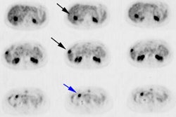
Results of preliminary clinical trials of the positron emission mammography
system PEM-I: a dedicated breast imaging system producing glucose metabolic
images using FDG.
Murthy K, Aznar M, Thompson CJ, Loutfi A, Lisbona R, Gagnon JH.
Early detection of breast cancer is crucial for efficient and effective
treatment. We have developed an instrument for positron emission mammography (PEM)
called PEM-I that performs high-resolution metabolic imaging of breast cancer.
Images of glucose metabolism are obtained after injection of 75 MBq FDG. The PEM
detectors are integrated into a conventional mammography system, allowing
acquisition of the emission images immediately after the mammogram, without
subject repositioning, and accurate coregistration of images from the 2
modalities. In this article, we present the results of the first clinical pilot
study with the instrument. METHODS: Sixteen subjects (age range, 34-76 y) were
studied. All subjects were nondiabetic, nonpregnant, and without a history of
cancer. They had recently been found to have suggestive mammography findings or
a palpable breast mass and underwent lumpectomy or mastectomy within 2 wk of the
study. Results from the PEM study were compared with those from mammography and
pathology. A PEM test was classified positive (indicating the presence of
cancer) if significant focal uptake was seen in the image or if the counting
rate in the breast with suggestive findings was significantly higher than in the
contralateral breast. RESULTS: Of the 16 subjects studied, 14 were evaluable.
Ten cancerous tumors and 4 benign tumors were confirmed by pathologic
examination after complete removal of the tumor. PEM correctly detected the
presence of disease in 8 of 10 subjects. Findings were false-negative in 2
instances and false-positive in none, giving the instrument 80% sensitivity,
100% specificity, and 86% accuracy. CONCLUSION: Our preliminary results suggest
that PEM can offer a noninvasive method for the diagnosis of breast cancer.
Metabolic images from PEM contain unique information not available from
conventional morphologic imaging techniques and aid in expeditiously
establishing the diagnosis of cancer. In all subjects, the PEM images were of
diagnostic quality, with an imaging time of 2-5 min.
PET > PET tumor imaging > Breast Cancer
J Nucl Med 2000 Nov;41(11):1851-8 (Comment in: J Nucl Med. 2000
Nov;41(11):1859-60)
Latest in PET
PET > PET tumor imaging > Neuroendocrine tumors
April 14, 2021
PET > PET tumor imaging > Head and Neck Tumors
March 12, 2018
PET > General > Images
January 23, 2018
PET > PET tumor imaging > Prostate cancer
January 20, 2016
