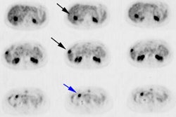
Evaluation of the (primary) tumor ('T')
- TX: cannot evaluate the primary tumor
- T0: no evidence of tumor
- T1:
tumor present, but not detectable clinically or with imaging
- T1a: tumor was incidentally found in less than 5% of prostate tissue resected (for other reasons)
- T1b: tumor was incidentally found in greater than 5% of prostate tissue resected
- T1c: tumor was found in a needle bniopsy performed due to an elevated serum PSA
- T2: the tumor can be felt (palpated) on examination,
but has not spread outside the prostate
- T2a: the tumor is in half or less than half of one of the prostate gland's two lobes
- T2b: the tumor is in more than half of one lobe, but not both
- T2c: the tumor is in both lobes but within the prostatic capsule
- T3: the tumor has spread through the prostatic capsule
(if it is only part-way through, it is still T2)
- T3a: the tumor has spread through the capsule on one or both sides
- T3b: the tumor has invaded one or both seminal vesicles
- T4: the tumor has invaded other nearby structures
It should be stressed that the designation "T2c" implies a tumor which is palpable in both lobes of the prostate. Tumors which are found to be bilateral on biopsy only but which are not palpable bilaterally should not be staged as T2c.
Evaluation of the regional lymph nodes ('N')
- NX: cannot evaluate the regional lymph nodes
- N0: there has been no spread to the regional lymph nodes
- N1: there has been spread to the regional lymph nodes
Evaluation of distant metastasis ('M')
- MX: cannot evaluate distant metastasis
- M0: there is no distant metastasis
- M1: there is distant metastasis
- M1a: the cancer has spread to lymph nodes beyond the regional ones
- M1b: the cancer has spread to bone
- M1c: the cancer has spread to other sites (regardless of bone involvement)
Evaluation of the histologic grade ('G')
Usually, the gradeof the cancer (how different the tissue is from normal tissue) is evaluated separately from the stage; however, for prostate cancer, grade information is used in conjunction with TNM status to group cases into four overall stages.
- GX: cannot assess grade
- G1: the tumor closely resembles normal tissue ( Gleason 2?4)
- G2: the tumor somewhat resembles normal tissue (Gleason 5?6)
- G3?4: the tumor resembles normal tissue barely or not at all (Gleason 7?10)
Of note, this system of describing tumors as "well-", "moderately-", and "poorly-" differentiated based on Gleason score of 2-4, 5-6, and 7-10, respectively, persists in SEER and other databases but is generally outdated. In recent years pathologists rarely assign a tumor a grade less than 3, particularly in biopsy tissue. A more contemporary consideration of Gleason grade is:
- Gleason 3+3: tumor is low grade (favorable prognosis)
- Gleason 3+4 / 3+5: tumor is mostly low grade with some high grade
- Gleason 4+3 / 5+3: tumor is mostly high grade with some low grade
- Gleason 4+4 / 4+5 / 5+4 / 5+5: tumor is all high grade
Note that under current guidelines, if any Pattern 5 is present it is included in final score, regardless of the percentage of the tissue having this pattern, as the presence of any pattern 5 is considered to be a poor prognostic marker.
Overall staging
The tumor, lymph node, metastasis, and grade status can be combined into four stages of worsening severity.
| Stage | Tumor | Nodes | Metastasis | Grade |
|---|---|---|---|---|
| Stage I | T1a | N0 | M0 | G1 |
| Stage II | T1a | N0 | M0 | G2?4 |
| T1b | N0 | M0 | Any G | |
| T1c | N0 | M0 | Any G | |
| T1 | N0 | M0 | Any G | |
| T2 | N0 | M0 | Any G | |
| Stage III | T3 | N0 | M0 | Any G |
| Stage IV | T4 | N0 | M0 | Any G |
| Any T | N1 | M0 | Any G | |
| Any T | Any N | M1 | Any G |
