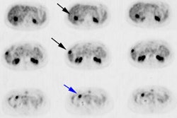
Breast cancer staging:
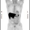
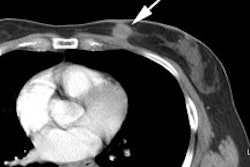
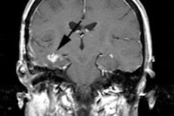
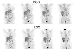
From J Nucl Med 2009; Lee JH, et al. The role of radiotracer imaging in the diagnosis and management of patients with breast cancer: part I- overview, detection, and staging. 50: 569-581
American Joint Committee on Cancer Staging System for Breast Cancer
| Tumor (T) | Definition |
| T0 | No primary tumor found |
| Tis | Carcinoma in situ |
| T1
T1mic T1a T1b T1c |
Invasive cancer less than 2cm in
greatest dimension
Tumor ≤ 0.1 cm in greatest dimension Tumor > 0.1 but ≤ 0.5 cm in greatest dimension Tumor > 0.5 cm but ≤ 1.0 cm in greatest dimension Tumor > 1.0 cm but ≤ 2.0 cm in greatest dimension |
| T2 | Tumor > 2 cm, but ≤ 5cm in greatest dimension |
| T3 | Tumor > 5 cm in greatest dimension |
| T4 | Tumor of any size with direct extension
to chest wall or skin T4a - extension to the chest wall T4b - ulceration, ipsilateral satellite nodules, and/or edema of the skin (including peau d'orange) T4c - T4a and T4b T4d - inflammatory carcinoma |
| Lymph nodes (N) | Definition |
| N0 | No regional lymph node mets |
| N1mi |
Micrometastases |
| N1 | Mets to mobile ipsilateral axillary level I-IInodes |
| N2 | Mets to fixed ipsilateral axillary nodes (N2a) or ipsilateral IM nodes in absence of ipsilateral axillary mets (N2b) |
| N3 | Mets to ipsilateral infraclavicular (level III) with or without axillary nodes (N3a); or ipsilateral IM and axillary nodes (N3b); or mets to ipsilateral supraclavicular nodes (N3c) |
M0 indicates no distant metastases, while M1 indicates distant mets
TNM staging system for invasive breast cancer
| Stage | Category |
| I | |
| IA |
T1N0M0 |
| IB |
T0N1mi (micrometastases)M0 T1N1miM0 |
| II | |
| IIA | T0N1M0
T1N1M0 T2N0M0 |
| IIB | T2N1M0
T3N0M0 |
| III | |
| IIIA | T3N1M0
T1-T3N2M0 |
| IIIB | T4N0-2M0 |
| IIIC | Any T N3M0 |
| IV | Any T, any N, M1 |
