Radiology 1999 Mar;210(3):807-14
Value of combined FDG PET and MR imaging in the evaluation of suspected
recurrent local-regional breast cancer: preliminary experience.
Hathaway PB, Mankoff DA, Maravilla KR, Austin-Seymour MM, Ellis GK, Gralow JR,
Cortese AA, Hayes CE, Moe RE.
PURPOSE: To assess the performance and potential clinical effects of combined
2-[fluorine 18]fluoro-2-deoxy-D-glucose (FDG) positron emission tomography (PET)
and magnetic resonance (MR) imaging of the axilla and brachial plexus in
patients suspected of having local-regional breast cancer metastases. MATERIALS
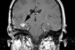
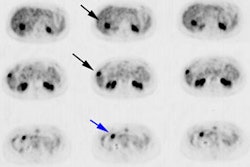
AND METHODS: Upper-body FDG PET and axillary and supraclavicular MR imaging were
performed in 10 patients (age range, 45-71 years) with clinical findings
suggestive of breast cancer metastases. Medical records were reviewed
retrospectively. Imaging findings were correlated with clinical data and
follow-up findings in all patients. Surgical findings were available in four
patients. RESULTS: Nine patients had local-regional breast cancer metastases. MR
imaging was diagnostic for tumor in five patients and was indeterminate in four
patients with axillary or chest wall metastases. With FDG PET, metastatic tumor
was positively identified in all nine patients. MR imaging was useful for
determining the relationship of metastatic tumor to axillary and supraclavicular
neurovascular structures. FDG PET helped confirm metastases in patients with
indeterminate MR imaging findings and depicted unsuspected metastases outside
the axilla. CONCLUSION: MR imaging and FDG PET are complementary in detecting
and characterizing local-regional breast cancer metastases. Combined FDG PET and
MR imaging provide useful treatment-planning data for patients clinically
suspected of having recurrent axillary or supraclavicular breast cancer.