Ann Oncol 1998 Oct;9(10):1117-22
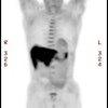
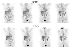
Whole-body 2-[18F]-fluoro-2-deoxy-D-glucose positron emission tomography (FDG-PET)
for accurate staging of Hodgkin's disease.
Bangerter M, Moog F, Buchmann I, Kotzerke J, Griesshammer M, Hafner M, Elsner K,
Frickhofen N, Reske SN, Bergmann L.
BACKGROUND: Staging of Hodgkin's disease (HD) is accomplished by a variety of
invasive and non-invasive modalities. This prospective study was undertaken to
investigate the value of whole-body positron emission tomography (PET) with
2-[18F]-fluoro-2-deoxy-D-glucose (FDG) in defining regions involved by lymphoma
compared with conventional staging methods in patients with HD. PATIENTS AND
METHODS: Fourty-four newly diagnosed patients with HD underwent FDG-PET as part
of their initial staging work-up. PET findings were correlated with findings of
conventional staging including computed tomography, ultrasound, bone scanning,
bone marrow biopsy, liver biopsy and laparotomy. When results of FDG-PET
differed to those obtained by conventional methods reevaluation was performed by
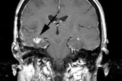
biopsy, if possible, or magnetic resonance imaging. RESULTS: The results of FDG-PET
were compared with three hundred twenty-one conventional staging procedures
performed in 44 patients. FDG-PET was positive in 38 of 44 (86%) patients at
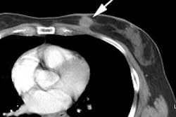
sites of documented disease. PET detected additional lesions in five cases
previously not identified by conventional staging methods. In another case a
nodal lesion suspect on CT was negative at FDG-PET and was settled as true
negative by biopsy. As a consequence of PET findings five patients had to be
upstaged and one patient had to be downstaged, resulting in changes in treatment
strategy in all six cases (14%). FDG-PET failed to visualize sites of HD in four
patients. In two of our patients a false positive PET result was obtained.
CONCLUSIONS: Our data indicate that FDG-PET provides an imaging technique that
appears to visualize involved lesions in most patients with HD and is useful in
the management of these patients.
