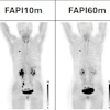
LOS ANGELES -- An investigational SPECT/CT radiotracer can accurately differentiate inflammation from fibrosis in interstitial lung disease (ILD), according to a study presented at the Society of Nuclear Medicine and Molecular Imaging 2026 Annual Meeting.
The finding is from a phase II trial involving 15 participants, with technitium-99m (Tc-99m) maraciclatide showing distinct uptake in ILD patients versus healthy controls, according to the researchers.
"While current imaging techniques can provide a structural view of fibrosis in the lungs, there is no reliable, noninvasive way to identify inflammation,” said study lead Druin Burch, MD, of John Radcliffe Hospital in Oxford, U.K., in an SNMMI news release. “A tool that could detect inflammation in ILD patients could help pinpoint those most likely to respond to anti-inflammatory therapy."
ILD includes more than 200 different types of lung conditions, and affects approximately 650,000 people in the U.S., resulting in an estimated 25,000 to 30,000 deaths per year. Differentiating between fibrotic scarring and inflammation stages of disease is critical so that physicians can determine what treatment is best for the patient.
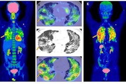
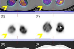
To assess whether Tc-99m maraciclatide SPECT/CT could play a role in these cases, Burch and colleagues administered 740 MBq of the tracer to five patients with idiopathic pulmonary fibrosis (IPF), five with fibrotic hypersensitivity pneumonitis (fHP), and five age- and sex-matched healthy controls. SPECT/CT scans were acquired two hours post-injection and evaluated by nuclear medicine physicians and thoracic radiologists based on radiological patterns, standardized uptake values, and target-to-background ratios.
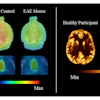
 Sample images from each participant group: A: CT only; B: fused Tc-99m maraciclatide SPECT/CT.Druin Burch, MD, and SNMMI
Ultimately, the results warrant a phase III study in a larger patient population, as is required before this imaging approach can be used outside the research setting, the researchers noted. As Tc‑99m maraciclatide has been granted fast-track designation by the U.S. Food and Drug Administration (FDA) for imaging ILD, it could become available to patients within two years of initiating a phase III trial, according to the group.
“Being able to differentiate the fibrotic and inflammation stages of ILD is not just beneficial to inform treatment decisions, but also for the development new therapies,” said Burch. “This approach has the potential to unlock a wide range of anti-inflammatory drugs for ILD.”
Check out AuntMinnie’s full coverage of SNMMI 2026 on our ShowCast.

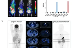


![(A-C) Representative whole-body maximum-intensity projection images and regional fused PET/CT images from three histologically confirmed osteosarcoma patients who underwent paired [68Ga]Ga-B7-H3-BCH PET/CT and 18F-FDGE PET/CT within seven days. (D) Multimodal imaging evaluation of Patient Three, including x-ray, MRI (T2-weighted imaging, T2WI), CT, and B7-H3 PET/CT.](https://img.auntminnie.com/mindful/smg/workspaces/default/uploads/2026/05/mei.XUQJWkpAJI.jpg?auto=format%2Ccompress&dpr=2&fit=crop&h=167&q=70&w=250)


![RET-targeted PET tracer highlights neuroendocrine prostate cancer tumors. Representative PET imaging shows strong tumor uptake of the RET-binding peptide tracer [⁶⁸Ga]Ga-DOTA-RET-L7 in a neuroendocrine prostate cancer (NEPC) model, supporting highly specific, high-contrast detection.](https://img.auntminnie.com/mindful/smg/workspaces/default/uploads/2026/05/screenshot-2026-05-27-205827.278Ys6PYU3.png?auto=format%2Ccompress&dpr=2&fit=crop&h=167&q=70&w=250)


