LOS ANGELES -- A new PET imaging approach for bone cancer can distinguish tumor tissue from normal tissue and assess surgical margins, according to research presented at the Society of Nuclear Medicine and Molecular Imaging (SNMMI) 2026 meeting.
The approach has the potential to reshape surgical practice for osteosarcoma by allowing for precise tumor resection -- which could significantly reduce the risk of local recurrence and preserve limb function, according to a team led by Bo Mei, PhD, of Peking University Cancer Hospital and Institute in Beijing.
"Despite continuous advances in surgical techniques, orthopedic surgeons still face a major challenge: how to delineate tumor margins accurately during surgery so as to ensure complete tumor removal while maximizing preservation of limb function," Mei said in a statement released by the society. "The development of an innovative technology capable of rapidly and reliably distinguishing tumor tissue from normal tissue and accurately assessing surgical margins in real time has become an urgent clinical priority in osteosarcoma management."
Osteosarcoma is among the most aggressive primary malignant bone tumors in children and adolescents, the group explained, noting that the current standard of care consists of chemotherapy combined with surgery. The main objective of surgical treatment is complete tumor removal, since positive margins increase the risk of local recurrence and affect long-term survival.
As high expression of the protein B7‑H3 is found in more than 80% of osteosarcoma cases, it's an attractive target for both imaging and therapy, the investigators wrote. For their research, they developed and synthesized a B7-H3-targeted radiotracer, 68Ga-B7H3-BCH, and conducted preclinical studies with five human osteosarcoma specimens to assess its ability to detect lesions in cell lines; single-cell RNA sequencing of the samples identified 13 cellular clusters within the tumor microenvironment. The team examined the specimens via PET imaging and a near-infrared B7H3 fluorescent probe.
The investigators reported the following:
- Malignant osteosarcoma cells were clearly distinguished from stromal and immune cells.
- Samples with high B7-H3 expression had worse event-free survival compared to the low-expression samples.
- Normal bone and a range of musculoskeletal tissues -- including periosteum, muscle, fascia, ligament, cartilage, nerve, growth plate, meniscus, bone marrow, and adipose tissue -- showed consistently low or negligible B7-H3 expression, which the team noted "confirmed excellent tumor specificity."
The study "establishes B7-H3 as a highly specific and clinically meaningful target in osteosarcoma and introduces a B7-H3–directed PET/NIR-II dual-modality imaging platform," the group wrote.
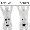
![(A-C) Representative whole-body maximum-intensity projection images and regional fused PET/CT images from three histologically confirmed osteosarcoma patients who underwent paired [68Ga]Ga-B7-H3-BCH PET/CT and 18F-FDGE PET/CT within seven days. (D) Multimodal imaging evaluation of Patient Three, including x-ray, MRI (T2-weighted imaging, T2WI), CT, and B7-H3 PET/CT.](https://img.auntminnie.com/mindful/smg/workspaces/default/uploads/2026/05/mei.XUQJWkpAJI.jpg?auto=format%2Ccompress&dpr=2&fit=max&q=70&w=700) (A-C) Representative whole-body maximum-intensity projection images and regional fused PET/CT images from three histologically confirmed osteosarcoma patients who underwent paired [68Ga]Ga-B7-H3-BCH PET/CT and 18F-FDGE PET/CT within seven days. (D) Multimodal imaging evaluation of Patient Three, including x-ray, MRI (T2-weighted imaging, T2WI), CT, and B7-H3 PET/CT.Bo Mei, PhD, and SNMMI
(A-C) Representative whole-body maximum-intensity projection images and regional fused PET/CT images from three histologically confirmed osteosarcoma patients who underwent paired [68Ga]Ga-B7-H3-BCH PET/CT and 18F-FDGE PET/CT within seven days. (D) Multimodal imaging evaluation of Patient Three, including x-ray, MRI (T2-weighted imaging, T2WI), CT, and B7-H3 PET/CT.Bo Mei, PhD, and SNMMI
"The development and clinical translation of this integrated platform will facilitate a paradigm shift in osteosarcoma care, from empirical 'surgery plus systemic chemotherapy' to individualized, precision, closed-loop diagnosis and treatment carrying major clinical and scientific significance," Mei said.
Check out AuntMinnie’s full coverage of SNMMI 2026 on our ShowCast.


![RET-targeted PET tracer highlights neuroendocrine prostate cancer tumors. Representative PET imaging shows strong tumor uptake of the RET-binding peptide tracer [⁶⁸Ga]Ga-DOTA-RET-L7 in a neuroendocrine prostate cancer (NEPC) model, supporting highly specific, high-contrast detection.](https://img.auntminnie.com/mindful/smg/workspaces/default/uploads/2026/05/screenshot-2026-05-27-205827.278Ys6PYU3.png?auto=format%2Ccompress&dpr=2&fit=crop&h=167&q=70&w=250)


