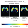
PET imaging shows lower levels of a receptor called muscarinic M1 across multiple brain regions in patients with schizophrenia, according to research delivered June 2 at the Society of Nuclear Medicine and Molecular Imaging (SNMMI) meeting.
The finding comes from the first in vivo PET study of M1 receptor availability in schizophrenia patients using the radiotracer carbon-11 (C-11) LSN3172176, with results supporting M1 as a treatment target, reported presenter Tommaso Volpi, MD, PhD, of Yale University, and colleagues.
"Our findings of lower muscarinic M1 receptor availability across several cortical and subcortical regions concur with postmortem data and support M1 as a target for [schizophrenia] treatment," the group wrote.
Muscarinic M1 is a receptor primarily located in the central nervous system, specifically in the cerebral cortex and hippocampus. It plays a crucial role in cognitive processing, learning, and memory. Postmortem studies strongly suggest that brain muscarinic M1 receptor deficits are present in a subset of schizophrenia patients, the researchers explained.
In September 2024, the U.S. Food and Drug Administration (FDA) approved Cobenfy, which contains the muscarinic M1/M4 agonist xanomeline, as the first non-dopaminergic medication for treating schizophrenia. While PET studies have confirmed that C-11 LSN3172176 binds with high affinity to muscarinic M1 in healthy controls, to date no studies have assessed the technique in patients with schizophrenia, the researchers noted.
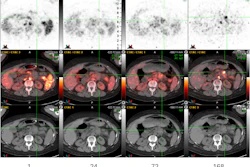
To bridge the gap, the Yale team recruited 16 schizophrenia patients (mean age 39 years old) and 16 age-matched healthy controls (mean age 37 years old) who underwent a two-hour PET scan with C-11 LSN3172176 on a high-resolution research tomograph system. M1 receptor binding was quantified using two methods, one requiring arterial blood sampling in a subset of participants, and one using a reference tissue approach (distribution volume ratio relative to the centrum semiovale [DVR(CS)]).
According to the analysis, schizophrenia patients showed significantly lower M1 binding than controls in the temporal, occipital, and parietal cortex, as well as the caudate, putamen, and amygdala. At the individual level, seven schizophrenia patients (44%) showed greater than 20% reduction in cortical DVR(CS) relative to the healthy controls, and 27% showed greater than 20% reduction in VT. No significant correlations were found between M1 binding and the Positive and Negative Syndrome Scale, a standard medical tool used to measure the symptom severity of schizophrenia, the group noted.
“This is the first in vivo PET imaging study of M1 receptor availability in schizophrenia using C-11 LSN3172176,” the group wrote.
Ultimately, the study confirms that deficits in M1 receptor are present in a subgroup of schizophrenia patients first described in 2024 as the “muscarinic receptor deficit intermediate phenotype,” according to the researchers.
Larger studies are warranted to examine correlations with clinical outcomes and treatment response to M1-targeting therapies, they concluded.
Check out AuntMinnie’s full coverage of SNMMI 2026 on our ShowCast.