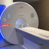
The U.S. Food and Drug Administration (FDA) has cleared Radialis' PET Imager, a small footprint, organ-targeted device.
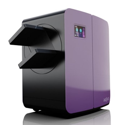
 The Radialis PET Imager. Image courtesy of Radialis.
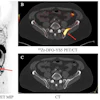
The system offers high spatial resolution and consists of a small field-of-view camera developed for close-range imaging, according to the firm. It features a partial-ring planar PET camera fitted with lutetium-based gamma-ray detectors that collect gamma rays emitted by radiopharmaceuticals injected into patients, the company said.
The technology is based on research that was conducted at Lakehead University in Thunder Bay, Ontario, Canada.