Having to retake MRI scans due to patient motion can easily cost a hospital upward of $100,000 in lost annual revenue, according to a study published in the July issue of the Journal of the American College of Radiology.
Analyzing a random week of MRI activity, researchers from the University of Washington in Seattle found that patient motion can be costly in terms of repeated sequences, which occurred in approximately 20% of MRI exams. A hospital or imaging center could lose $140,000 or more in annual revenue per scanner based on that rate of repeat exams, they concluded (JACR, July 2015, Vol. 12:7, pp. 689-605).
The findings could prompt the development of more advanced technology and software to correct for patient motion, the authors added. Such technology would also benefit radiologists who have to read blurry MRI scans.
"We, as radiologists, tend to read through motion if it is not too severe. We see it fairly frequently, and most of the time we can get by," lead author Dr. Jalal Andre, an assistant professor of radiology, told AuntMinnie.com. "But I wonder how much longer we are going to tolerate artifacts from patient motion in reading exams."
Previous research
Past studies have estimated that patient motion adversely affects MR image quality in 10% to 42% of sequences. Motion artifacts may be even more prevalent in advanced MRI techniques that rely on higher spatial resolution.
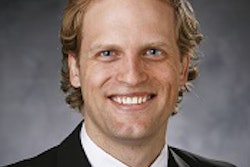
 Dr. Jalal Andre from the University of Washington.
"Surprisingly, the prevalence and severity of motion artifacts that result in significantly degraded clinical MR exams is poorly documented in the literature, as is their impact on throughput and organizational finances," Andre and colleagues wrote.
Their retrospective study reviewed clinical MRI exams in one randomly selected week in April 2014. The exams were reviewed by neuroradiologists who were blinded to medical history and clinical presentation, as well as to whether the scans were acquired in an outpatient, inpatient, or emergency department (ED) setting.
The MRI exams were performed on three different scanners (1.5-tesla Avanto and 3-tesla Tim Trio, Siemens Healthcare; 1.5-tesla Signa HD, GE Healthcare), all of which featured a conventional closed configuration with a 60-cm bore.
A total of 192 clinical MRI exams were included in the study. The patient population consisted of 97 women and 95 men between the ages of 21 and 87 years (mean age, 56 years).
In terms of setting, 118 exams were performed on an outpatient basis and 76 were inpatient and/or ED exams. Also, of the 192 exams, 173 (90%) were limited to the neuroaxis and 120 (63%) were limited to the brain and head and neck region.
Monetary calculations
Dollar amounts were calculated based on the cost of a routine noncontrast-enhanced MRI brain scan using the CPT code 70551 for outpatient costs. For inpatient costs, the researchers added institutional operational-dollar estimates for MRI and relative value units (RVUs) per CPT codes and patient volumes.
Andre and colleagues assumed 45-minute time slots for initial MRI scans and used the actual scan times for repeat sequences, taking the data directly from the scanner's console.
Thus, the base-case cost estimate for a brain MRI exam was $444.32 for a 45-minutes time slot, which was extrapolated to a cost of $592 per hour. This value was then used to estimate the expense of repeat sequences associated with motion artifacts.
The researchers did not take into account differences in reimbursement in other parts of the U.S. for scanning in the outpatient, inpatient, and/or ED setting, nor did they include variations in technical and/or professional fees.
However, they did apply a 20% deviation to their hourly base-case cost scenario to provide a range of possible financial implications from patient motion and to expand the potential of lost revenue, from a low estimate of $474 per hour to a high estimate of $711 per hour.
"If you think about cost as a penalty in terms of patient motion, the penalty will change based on reimbursement rates and where you are in the country, because reimbursement rates fluctuate based on geographic location," Andre said.
Motion prevalence
Among the 192 exams evaluated, 203 sequences had moderate and/or severe motion artifacts and suboptimal image quality. Sixty-eight of the subpar MRI sequences were repeated; these came from 38 patients.
A breakdown of patient groups found significant motion artifacts on MRI sequences in 29% of inpatient and/or emergency department exams, compared with only 7% of outpatient MRI scans.
Andre said he and his colleagues were not surprised to find that inpatients and ED patients tended to move more often than outpatients during MRI scans, but they did not expect such a significant disparity between the two groups.
"We also were surprised to see how often outpatients moved, because they are supposedly relatively healthy and can prepare themselves for the scan," he said. "Having 7% of outpatients with minimal and moderate motion was somewhat surprising."
The repeating of 68 MRI sequences required 278.5 minutes of additional scan time to produce adequate image quality. Based on the study's average base-case MRI cost of $592 per hour, the extra scanning time that week cost the hospital $2,750 in lost revenue.
When the results for that week are extrapolated to a full year, the hospital could lose more than $140,000 annually in revenue and/or additional costs, Andre and colleagues estimated. When they added the 20% deviation variable, the estimated amount for nonreimbursed MR scans ranged from $114,000 to $172,000.
If all 203 sequences found to contain moderate and/or severe motion artifacts were counted, this would represent lost revenue of approximately $8,351 per week or $434,000 per year. Adding the 20% deviation variable resulted in a range of $347,000 to $521,000 per year.
"This is a small study, and we need to expand the evaluation time frame and the body parts that were evaluated," Andre said. "Looking into the future, the evaluation of motion that includes respiratory and cardiac motion, which basically is core body motion, would be of value, as well as expanding the study population."