NEW ORLEANS – Post-therapy SPECT/CT is invaluable for patients undergoing radiopharmaceutical therapy, according to a June 21 presentation Society of Nuclear Medicine and Molecular Imaging 2025 annual meeting.
In a case series, the method significantly enhanced patient safety, optimized therapy regimens, and improved outcomes for patients, noted presenter Taylor Gillespie, a nuclear medicine clinical research leader at the University of Tennessee in Knoxville.
“Most institutions only look at imaging and dosimetry to adjust a patient’s dose rather than to assess the impact it can have on overall care,” Gillespie said. “In our program, we perform post-therapy imaging and dosimetry for every patient receiving lutetium-based therapies and have seen a significant enhancement in patient management.”
Radiopharmaceutical therapy is rapidly gaining momentum in the U.S., fueled by recent approvals that expand access to targeted cancer treatments. However, post-therapy imaging and dosimetry -- long considered essential in external beam radiation oncology -- have yet to become standard practice in the field, Gillespie noted.
To illustrate its value, Gillespie presented seven case reports from patients who received standard doses of either lutetium-177 (Lu-177) DOTATATE (Lutathera, Novartis) or Lu-177 prostate-specific membrane antigen (PSMA) (Pluvicto, Novartis), followed by post-therapy SPECT/CT imaging at two to four hours and five to seven days postinfusion. Using advanced software, technologists and physicists measured how long the radiation remained in different organs and calculated the radiation dose each organ received.
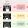
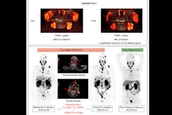
 Top image displays a patient assessed for acute renal failure following third Lutathera administration where 3D dosimetry showed an average-per-administration kidney absorbed dose of 5 Gray, with total course of therapy being well under toxicity limits. Bottom image displays a patient’s absorbed dose to the colon after his first dose of Pluvicto where 3D dosimetry was used to confirm transient bowel obstruction leading to treatment management.Taylor Gillespie and SNMMI
The imaging helped identify causes of complications, such as noncompliance with hydration guidelines, radiation exposure to sensitive organs, and unexpected disease findings. In addition, dosimetry-guided clinical decisions to continue, modify, or stop therapy prompted changes in imaging protocols, improved symptom management, and enhanced radiation safety measures, Gillespie reported.
Ultimately, post-therapy imaging optimized treatment outcomes and protected patients, he said.
“These cases show that post-therapy imaging and dosimetry -- instead of just following generic rules or assumptions -- can make treatment safer and personalized. If more hospitals conduct post-therapy imaging and dosimetry, it could lead to national guidelines that help patients everywhere, even in places without specialized staff,” Gillespie concluded.