Neurofibroma:
Clinical:
Neurofibromas account for only 10% of mediastinal neurogenic tumors. Most patients present between the ages of 20-40 years [3]. Neurofibromas are rare in children. About 30-45% of patients have neurofibromatosis (NF1). It is a mixed tumor containing both Schwann cells and nerve cells, and the tumor is not separated from the parent nerve. Areas of cystic degeneration, hypocellularity, and xanthomatous material are uncommon [3]. Resection is curative.In cases where the tumor creates a thick, convoluted mass that invovles multiple fascicles of a nerve and infiltrates along an entire nerve trunk it is termed a "plexiform neurofibroma"- these are considered pathognomonic of Neurofibromatosis Type I [3]. Although plexiform neurofibromas are considered benign, they carry a risk of being malignant in 2-5% of cases [3].
X-ray:
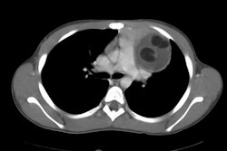
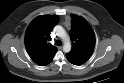
On CT they appear as sharply marginated, homogeneous, slightly low density soft tissue masses which may cause pressure erosion of adjacent bone or appear as a dumb-bell lesion. Calcification is rare. There is homogeneous enhancement following contrast administration [3]. It is often difficult to distinguish from a schwannoma in its appearance.On MR, the lesion has homogeneous low to intermediate intensity on T1 images [3]. On T2 images, the peripheral zone sometimes shows higher signal intensity than the central zone [3].
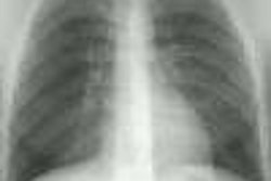
A plexiform neurofibroma is a large mass that may encase a rib producing a "ribbon rib". This lesion is usually seen in patients with neurofibromatosis type I and may degenerate to form a malignant lesion.
REFERENCES:
(1) Chest 1997; Strollo DC,
et
al. Primary mediastinal tumors. Part II: Tumors of the
middle and posterior
mediastinum. 112: 1344-57
(2) AJR 1999; Rossi SE, et
al.
Thoracic manifestations of Neurofibromatosis-I. 173: 1631-1638
(3) AJR 2016; Pavlus JD, et al. Imaging of thoracic neurogenic tumors. 207: 552-561